
Page 58 - Read Online
P. 58
Malcher et al. Mini-invasive Surg 2021;5:31 https://dx.doi.org/10.20517/2574-1225.2021.48 Page 5 of 8
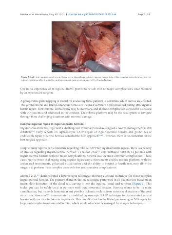
Figure 2. Right side inguinoscrotal hernia. Green circle shows large indirect inguinal hernia defect. Black arrows show distal edge of the
indirect hernia sac after transection and blue arrows show proximal edge of the transected sac.
Our initial experience of 10 inguinal RoME proved to be safe with no major complications, once executed
by an experienced surgeon.
A preoperative pain mapping is crucial for evaluating these patients to determine which nerves are affected.
The genitofemoral and lateral-cutaneous nerves are the most common nerves involved during MIS inguinal
hernia repair. Furthermore, orchiectomy may be necessary, and all these complications should be discussed
with the patients and addressed in the consent. The robotic platform may be the best option to navigate
through these challenging situations with minimal damage.
Robotic inguinal repair in inguinoscrotal hernias
Inguinoscrotal hernias represent a challenge for minimally invasive surgeons, and its management is still
[38]
debatable . Early reports on laparoscopic TAPP repair of inguinoscrotal hernias and guidelines of
endoscopic repair of scrotal hernias validated the MIS approach [39,40] . However, there is no consensus on the
best surgical approach.
Despite many reports in the literature regarding robotic TAPP for inguinal hernia repairs, there is a paucity
[41]
of studies regarding inguinoscrotal hernias . Yheulon et al. demonstrated rIHR in 14 patients with
[41]
inguinoscrotal hernias with no major complications. Seroma was the most common complication. These
cases may be more challenging using regular laparoscopic instruments and the robotic platform, with the
articulated instruments, enhanced visualization and the ability to control a fourth arm, may allow the
surgeon to perform these complex cases with few post-operative complications.
Morrell et al. demonstrated a laparoscopic technique showing a special technique for those complex
[38]
inguinoscrotal hernias. The primary abandon-the-sac technique performed in 26 patients was based on an
incomplete dissection of the distal sac, leaving it into the inguinal canal and scrotum [Figure 2]. This
technique can be safely used in patients with inguinoscrotal hernias. Seroma seems to be its main
complication, but it avoids hematomas and possible ischemic orchitis from extensive dissection of the cord
[42]
structures. Siow et al. demonstrated a modified laparoscopic TAPP technique for incarcerated scrotal
hernias with a scrotal incision in 20 patients. This modification has facilitated performing an MIS repair for
large and complex inguinoscrotal hernias, which would otherwise be managed by an open technique.