
Page 56 - Read Online
P. 56
Malcher et al. Mini-invasive Surg 2021;5:31 https://dx.doi.org/10.20517/2574-1225.2021.48 Page 3 of 8
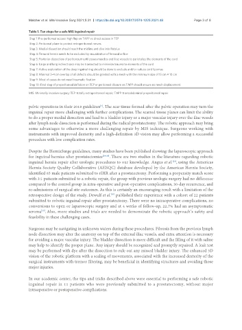
Table 1. Ten steps for a safe MIS inguinal repair
Step 1: Pre-peritoneal access: high flap on TAPP vs direct access in TEP
Step 2: Peritoneal plane to protect retroperitoneal nerves
Step 3: Medial dissection should reach the midline and dive into Retzius
Step 4: Femoral hernia needs to be excluded by visualization of femoral orifice
Step 5: Posterior dissection of peritoneum until psoas muscles and iliac vessels to parietalize the elements of the cord
Step 6: Large and long indirect sacs may be transected to minimize trauma to elements of the cord
Step 7: Active exploration of the deep inguinal ring should be done to exclude and/or reduce cord lipomas
Step 8: Minimal 3-4 cm overlap of all defects should be granted with a mesh with the minimum size of 15 cm × 10 cm
Step 9: Most of cases do not need traumatic fixation
Step 10: Final step of preperitoneal deflation on TEP or peritoneal closure on TAPP should ensure no mesh displacement
MIS: Minimally invasive surgery; TEP: totally extraperitoneal repair; TAPP: transabdominal preperitoneal repair.
pelvic operations in their 2018 guidelines . The scar tissue formed after the pelvic operation may turn the
[5]
inguinal repair more challenging with further complications. The scarred tissue planes can limit the ability
to do a proper medial dissection and lead to a bladder injury or a major vascular injury over the iliac vessels
after lymph node dissection is performed during the radical prostatectomy. The robotic approach may bring
some advantages to otherwise a more challenging repair by MIS technique. Surgeons working with
instruments with improved dexterity and a high-definition 3D vision may allow performing a successful
procedure with low complication rates.
Despite the HerniaSurge guidelines, many studies have been published showing the laparoscopic approach
for inguinal hernias after prostatectomies [24-28] . There are two studies in the literature regarding robotic
inguinal hernia repair after urologic procedures to our knowledge. Angus et al. , using the Americas
[29]
Hernia Society Quality Collaborative (AHSQC) database developed by the Americas Hernia Society,
identified 65 male patients submitted to rIHR after a prostatectomy. Performing a propensity match score
with 3:1 patients submitted to a robotic repair, the group with previous urologic surgery had no difference
compared to the control group in intra-operative and post-operative complications, 30-day recurrence, and
re-admissions of surgical site outcomes. As this is certainly an encouraging result with a limitation of the
[30]
retrospective design of the study. Dewulf et al. published their experience with a cohort of 22 patients
submitted to robotic inguinal repair after prostatectomy. There were no intraoperative complications, no
conversions to open or laparoscopic surgery and at 4 weeks of follow-up, 22.7% had an asymptomatic
seroma . Also, more studies and trials are needed to demonstrate the robotic approach’s safety and
[30]
feasibility in these challenging cases.
Surgeons may be navigating in unknown waters during these procedures. Fibrosis from the previous lymph
node dissection may alter the anatomy on top of the external iliac vessels, and extra attention is necessary
for avoiding a major vascular injury. The bladder dissection is more difficult and the filling of it with saline
may help to identify the proper plane. Any injury should be recognized and promptly repaired. A leak test
may be performed with dye after the dissection to rule out any missed bladder injury. The enhanced 3D
vision of the robotic platform with a scaling of movements, associated with the increased dexterity of the
surgical instruments with tremor filtering, may be beneficial in identifying structures and avoiding those
major injuries.
In our academic center, the tips and tricks described above were essential to performing a safe robotic
inguinal repair in 11 patients who were previously submitted to a prostatectomy, without major
intraoperative or postoperative complications.