
Page 43 - Read Online
P. 43
Baur et al. Mini-invasive Surg 2021;5:27 https://dx.doi.org/10.20517/2574-1225.2021.28 Page 5 of 7
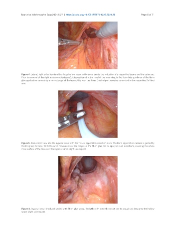
Figure 4. Lateral, right sided hernia with a large hollow space in the deep, due to the reduction of a respective lipoma and the outer sac.
Prior to removal of the right instrument (scissors), it is positioned at the level of the inner ring, to facilitate later guidance of the fibrin
glue application cannula by a correct angel of the trocar; this way, the 8 mm DaVinci port remains connected to the respective DaVinci
arm.
Figure 5. Endoscopic view into the inguinal canal with the Tisseel applicator already in place. The fibrin application cannula is guided by
the Prograsp forceps. With the wrist movements of the Prograsp, the fibrin glue can be sprayed in all directions, covering the whole
inner surface of the tissues of the inguinal canal (right side repair).
Figure 6. Inguinal canal lined and sealed with fibrin glue spray. With the 30° optic the result can be visualised deep into the hollow
space (right side repair).