
Page 42 - Read Online
P. 42
Page 4 of 7 Baur et al. Mini-invasive Surg 2021;5:27 https://dx.doi.org/10.20517/2574-1225.2021.28
Figure 1. Initial situation with a right sided large direct hernia (EHS M3).
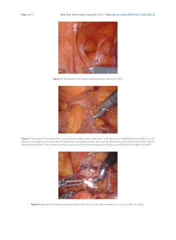
Figure 2. The suture of transverse fascia is performed step-by-step in alternation with the gradual mobilisation of the fatty tissue. In
doing so, the surgeon can be sure that the suture does not damage vessels, nerves or the deferent duct, structures that are very close to
the fascia transversalis. This prevents excessive protrusion of the transverse fascia into the groin during dissection (right side repair).
Figure 3. Suturing of the transverse fascia down to the iliopubic tract with a running V-Loc suture (right side repair).