
Page 34 - Read Online
P. 34
Page 6 of 9 Okano et al. Mini-invasive Surg 2021;5:29 https://dx.doi.org/10.20517/2574-1225.2021.15
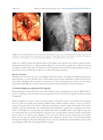
Figure 4. Case of papillary perforation after endoscopic sphincterotomy. The bile duct (arrowhead) and the perforation (arrow) were
confirmed. Covered metallic stent was placed for the purpose of compression closure, and endoscopic nasobiliary drainage was
performed. It subsequently conservatively alleviated the complication, and surgical treatment was avoided.
guide wire could be passed through the inside of the plastic stent, and the stent could be removed with a
Soehendra Stent Retriever or a balloon catheter [Figure 5]. In cases where a guide wire could not be passed
®
through the inside of the stent, the distal end could be grasped with a basket forceps, grasping forceps, or
polypectomy snare, and the stent could recover under fluoroscopic guidance.
Bile duct bleeding
Bleeding from the tumor may occur in malignant bile duct stricture. Although this bleeding usually stops
spontaneously, a covered metallic stent could be placed and pressure hemostasis could be performed in
cases where bleeding from the tumor is continuous and the anemia progresses (video). In cases where
hemostasis is still difficult to achieve, hemostasis by interventional radiology should be considered.
LITHIASIS REMOVAL-ASSOCIATED ISSUES
When grasping a common bile duct stone with a basket forceps, crushing the stone may be difficult due to
its size or hardness, and the basket may be impossible to pull out from the papilla, thereby becoming
strangulated.
When the papillary incision is small, the outside sheath of the basket forceps can be pulled off, leaving just
the wire. Then, the papilla can be further dilated using a dilation balloon catheter, and it can then be
removed along with the stone. If that is difficult, an endotripter may be useful. The handle of the basket
catheter is cut off, and thereafter, the metal sheath of the endotripter is passed after covering the wire of the
basket. In through-the-scope type thin endotripters, this action is possible without removing the scope, but
for endotripters that are not through-the-scope type, the scope should be removed, and the metal sheath
passed under X-ray fluoroscopic guidance. Once the metal sheath has been passed up to the basket
impaction, the wire can be fixed onto the handle of the endotripter, and the stone can be crushed releasing
the impaction [Figure 6]. Because basket impaction is a serious complication, it is important to always have
an endotripter ready while performing lithiasis treatment.