
Page 25 - Read Online
P. 25
Page 6 of 9 Aabakken et al. Mini-invasive Surg 2021;5:25 https://dx.doi.org/10.20517/2574-1225.2021.09
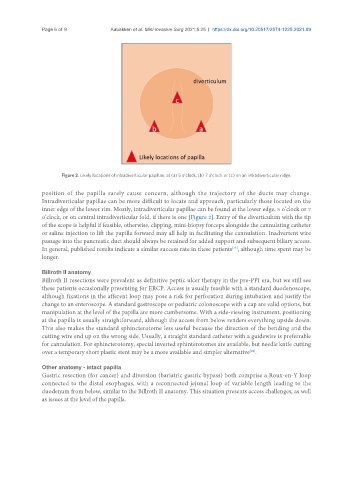
Figure 2. Likely locations of intradiverticular papillae, at (a) 5 o’clock, (b) 7 o’clock or (c) on an intradiverticular ridge.
position of the papilla rarely cause concern, although the trajectory of the ducts may change.
Intradiverticular papillae can be more difficult to locate and approach, particularly those located on the
inner edge of the lower rim. Mostly, intradiverticular papillae can be found at the lower edge, 5 o’clock or 7
o’clock, or on central intradiverticular fold, if there is one [Figure 2]. Entry of the diverticulum with the tip
of the scope is helpful if feasible, otherwise, clipping, mini-biopsy forceps alongside the cannulating catheter
or saline injection to lift the papilla forward may all help in facilitating the cannulation. Inadvertent wire
passage into the pancreatic duct should always be retained for added support and subsequent biliary access.
[23]
In general, published results indicate a similar success rate in these patients , although time spent may be
longer.
Billroth II anatomy
Billroth II resections were prevalent as definitive peptic ulcer therapy in the pre-PPI era, but we still see
these patients occasionally presenting for ERCP. Access is usually feasible with a standard duodenoscope,
although fixations in the afferent loop may pose a risk for perforation during intubation and justify the
change to an enteroscope. A standard gastroscope or pediatric colonoscope with a cap are valid options, but
manipulation at the level of the papilla are more cumbersome. With a side-viewing instrument, positioning
at the papilla is usually straightforward, although the access from below renders everything upside down.
This also makes the standard sphincterotome less useful because the direction of the bending and the
cutting wire end up on the wrong side. Usually, a straight standard catheter with a guidewire is preferrable
for cannulation. For sphincterotomy, special inverted sphinterotomes are available, but needle knife cutting
[24]
over a temporary short plastic stent may be a more available and simpler alternative .
Other anatomy - intact papilla
Gastric resection (for cancer) and diversion (bariatric gastric bypass) both comprise a Roux-en-Y loop
connected to the distal esophagus, with a reconnected jejunal loop of variable length leading to the
duodenum from below, similar to the Billroth II anatomy. This situation presents access challenges, as well
as issues at the level of the papilla.