
Page 22 - Read Online
P. 22
Aabakken et al. Mini-invasive Surg 2021;5:25 https://dx.doi.org/10.20517/2574-1225.2021.09 Page 3 of 9
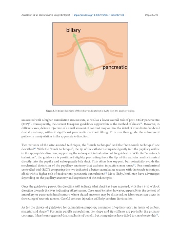
Figure 1. Principal directions of the biliary and pancreatic ducts from the papillary orifice.
associated with a higher cannulation success rate, as well as a lower overall risk of post-ERCP pancreatitis
[4]
(PEP) . Consequently, the current European guidelines support this as the method of choice . However, in
[2]
difficult cases, delicate injection of a small amount of contrast may outline the detail of mural intraduodenal
ductal anatomy, without significant pancreatic contrast filling. This can then guide the subsequent
guidewire manipulation in the appropriate direction.
Two variants of the wire-assisted technique, the “touch technique” and the “non-touch technique” are
described . With the “touch technique”, the tip of the catheter is impacted gently into the papillary orifice
[4]
in the appropriate direction, supporting the subsequent introduction of the guidewire. With the “non-touch
technique”, the guidewire is positioned slightly protruding from the tip of the catheter and is inserted
directly into the papilla and subsequently bile duct. This offers less support, but potentially avoids the
mechanical distortion of the papillary anatomy that catheter impaction may cause . One randomized
[5]
controlled trial (RCT) comparing the two indicated a better cannulation success with the touch-technique,
albeit with a higher risk of inadvertent pancreatic cannulations . Most likely, both may have advantages
[6]
depending on the papillary anatomy and experience of the endoscopist.
Once the guidewire passes, the direction will indicate what duct has been accessed, with the 11-12 o’clock
direction towards the liver indicating biliary access. Care must be taken however, especially in the context of
ampullary or pancreatic head tumors, where ductal anatomy may be distorted, or false routes can occur in
the setting of necrotic tumors. Careful contrast injection will help confirm the situation.
As for the choice of guidewire for cannulation purposes, a number of options exist, in terms of caliber,
material and shape . For main papilla cannulation, the shape and tip stiffness are probably the primary
[4]
concerns. It has been suggested that smaller is of benefit, but comparisons have failed to corroborate this ,
[7]