
Page 8 - Read Online
P. 8
Kauffmann et al. Mini-invasive Surg 2020;4:54 I http://dx.doi.org/10.20517/2574-1225.2020.46 Page 3 of 10
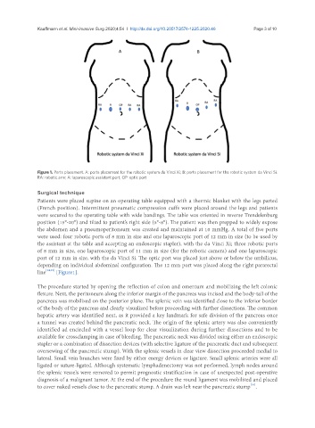
Figure 1. Ports placement. A: ports placement for the robotic system da Vinci Xi; B: ports placement for the robotic system da Vinci Si.
RA: robotic arm; A: laparoscopic assistant port; OP: optic port
Surgical technique
Patients were placed supine on an operating table equipped with a thermic blanket with the legs parted
(French position). Intermittent pneumatic compression cuffs were placed around the legs and patients
were secured to the operating table with wide bandings. The table was oriented in reverse Trendelenburg
position (15°-20°) and tilted to patient’s right side (5°-8°). The patient was then prepped to widely expose
the abdomen and a pneumoperitoneum was created and maintained at 10 mmHg. A total of five ports
were used: four robotic ports of 8 mm in size and one laparoscopic port of 12 mm in size (to be used by
the assistant at the table and accepting an endoscopic stapler), with the da Vinci Xi; three robotic ports
of 8 mm in size, one laparoscopic port of 11 mm in size (for the robotic camera) and one laparoscopic
port of 12 mm in size, with the da Vinci Si. The optic port was placed just above or below the umbilicus,
depending on individual abdominal configuration. The 12 mm port was placed along the right pararectal
line [14,15] [Figure1].
The procedure started by opening the reflection of colon and omentum and mobilizing the left colonic
flexure. Next, the peritoneum along the inferior margin of the pancreas was incised and the body-tail of the
pancreas was mobilized on the posterior plane. The splenic vein was identified close to the inferior border
of the body of the pancreas and clearly visualized before proceeding with further dissections. The common
hepatic artery was identified next, as it provided a key landmark for safe division of the pancreas once
a tunnel was created behind the pancreatic neck. The origin of the splenic artery was also conveniently
identified ad encircled with a vessel loop for clear visualization during further dissections and to be
available for crossclamping in case of bleeding. The pancreatic neck was divided using either an endoscopic
stapler or a combination of dissection devices (with selective ligature of the pancreatic duct and subsequent
oversewing of the pancreatic stump). With the splenic vessels in clear view dissection proceeded medial to
lateral. Small vein branches were fixed by either energy devices or ligature. Small splenic arteries were all
ligated or suture-ligated. Although systematic lymphadenectomy was not performed, lymph nodes around
the splenic vessels were removed to permit prognostic stratification in case of unexpected post-operative
diagnosis of a malignant tumor. At the end of the procedure the round ligament was mobilized and placed
[16]
to cover naked vessels close to the pancreatic stump. A drain was left near the pancreatic stump .