
Page 124 - Read Online
P. 124
Page 2 of 19 Gharagozloo et al. Mini-invasive Surg 2020;4:48 I http://dx.doi.org/10.20517/2574-1225.2020.35
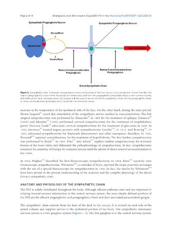
Figure 1. Sympathetic chain. Schematic representation shows the location of the first neuron in the spinal cord. Axons from the first
neuron (preganglionic) travel within the dorsal and ventral roots and form the preganglionic sympathetic fibers or rami communicantes
albi (white rami). Rami communicantes albi synapse with the second neuron within the sympathetic chain and the postganglionic fibers
or ramus communicantes grisei (grey rami) travel with the intercostal nerves
increase in the temperature of the ipsolateral side of the face. On the other hand, during the same period,
[4]
Brown-Sequard noted that stimulation of the sympathetic nerves resulted in vasoconstriction. The first
[5]
[6]
surgical sympathectomy was performed by Alexander in 1889 for the treatment of epilepsy. Jonnesco
[7]
(1896) and Jaboulay (1900) performed cervical sympathectomy for the treatment of exophthalmic
goiter. Francois-Frank advocated cervical sympathectomy for the treatment of glaucoma in 1899. In
[8]
[9]
[10]
[11]
1920, Jonnesco treated angina pectoris with sympathectomy. Leriche , in 1913, and Bruning , in
1923, advocated sympathectomy for Raynaud’s phenomenon and other vasospastic disorders. In 1920,
[12]
Kotzareff reported sympathectomy for the treatment of hyperhidrosis. The first lumbar sympathectomy
[15]
was performed by Royle in 1923. Diez and Adson applied lumbar sympathectomy for ischemic
[14]
[13]
lesions of the lower limbs and delineated the pathophysiology of sympathectomy. In fact, sympathectomy
remained the mainstay of therapy for ischemic lesions until the advent of direct arterial revascularization in
the 1960s.
[16]
[17]
In 1942, Hughes described the first thoracoscopic sympathectomy. In 1954, Krux reported 1400
[18]
thoracoscopic sympathectomies. Wittmoser , a coworker of Krux, reported the single-puncture technique
[19]
with the use of a special thoracoscope for sympathectomy in 1950. In fact, the studies by Wittmoser
have been pivotal in the present understanding of the anatomy and the complex physiology of the dorsal
thoracic sympathetic chain.
ANATOMY AND PHYSIOLOGY OF THE SYMPATHETIC CHAIN
The SNS is widely distributed throughout the body. Although afferent pathways exist and are important in
relaying visceral sensory information to the central nervous system, the most clearly defined portions of
the SNS are the efferent preganglionic and postganglionic fibers and their associated paravertebral ganglia.
The sympathetic chain extends from the base of the skull to the coccyx. It is located on each side of the
spinal column and supplies nerves to the ipsilateral portion of the body. The sympathetic autonomic
nervous system is a two-ganglion system [Figures 1-3]. The first ganglion is in the central nervous system,