
Page 11 - Read Online
P. 11
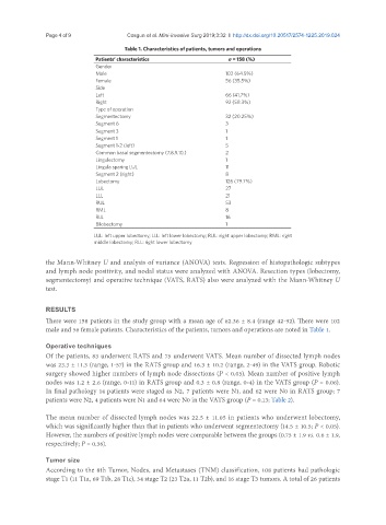
Page 4 of 9                                        Cosgun et al. Mini-invasive Surg 2019;3:32  I  http://dx.doi.org/10.20517/2574-1225.2019.024

                                       Table 1. Characteristics of patients, tumors and operations
                               Patients’ characteristics               n = 158 (%)
                               Gender
                               Male                                    102 (64.5%)
                               Female                                  56 (35.5%)
                               Side
                               Left                                    66 (41.7%)
                               Right                                   92 (58.3%)
                               Type of operation
                               Segmentectomy                           32 (20.25%)
                               Segment 6                               3
                               Segment 3                               1
                               Segment 1                               1
                               Segment 1-2 (left)                      5
                               Common basal segmentectomy (7.8.9.10.)  2
                               Lingulectomy                            1
                               Lingula sparing LUL                     11
                               Segment 2 (right)                       8
                               Lobectomy                               126 (79.7%)
                               LUL                                     27
                               LLL                                     21
                               RUL                                     53
                               RML                                     8
                               RLL                                     16
                               Bilobectomy                             1

                              LUL: left upper lobectomy; LLL: left lower lobectomy; RUL: right upper lobectomy; RML: right
                              middle lobectomy; RLL: right lower lobectomy

               the Mann-Whitney U and analysis of variance (ANOVA) tests. Regression of histopathologic subtypes
               and lymph node positivity, and nodal status were analyzed with ANOVA. Resection types (lobectomy,
               segmentectomy) and operative technique (VATS, RATS) also were analyzed with the Mann-Whitney U
               test.


               RESULTS
               There were 158 patients in the study group with a mean age of 62.36 ± 8.4 (range 42-92). There were 102
               male and 56 female patients. Characteristics of the patients, tumors and operations are noted in Table 1.


               Operative techniques
               Of the patients, 83 underwent RATS and 75 underwent VATS. Mean number of dissected lymph nodes
               was 23.3 ± 11.3 (range, 1-57) in the RATS group and 16.3 ± 10.2 (range, 2-49) in the VATS group. Robotic
               surgery showed higher numbers of lymph node dissections (P < 0.05). Mean number of positive lymph
               nodes was 1.2 ± 2.6 (range, 0-11) in RATS group and 0.3 ± 0.8 (range, 0-4) in the VATS group (P = 0.06).
               In final pathology 14 patients were staged as N2, 7 patients were N1, and 62 were N0 in RATS group; 7
               patients were N2, 4 patients were N1 and 64 were N0 in the VATS group (P = 0.13; Table 2).

               The mean number of dissected lymph nodes was 22.5 ± 11.05 in patients who underwent lobectomy,
               which was significantly higher than that in patients who underwent segmentectomy (14.5 ± 10.5; P < 0.05).
               However, the numbers of positive lymph nodes were comparable between the groups (0.75 ± 1.9 vs. 0.6 ± 1.9,
               respectively; P = 0.36).


               Tumor size
               According to the 8th Tumor, Nodes, and Metastases (TNM) classification, 108 patients had pathologic
               stage T1 (11 T1a, 69 T1b, 28 T1c), 34 stage T2 (23 T2a, 11 T2b), and 16 stage T3 tumors. A total of 26 patients