
Page 34 - Read Online
P. 34
Page 2 of 6 Zirafa et al. Mini-invasive Surg 2020;4:13 I http://dx.doi.org/10.20517/2574-1225.2019.35
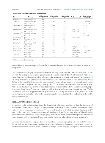
Table 1. Nodal upstaging in non-small cell lung cancer
Nodal upstaging N1 upstaging N2 upstaging Nodal stations
Ref. Patients Nodes removed
(%) (%) (%) examined
Rocha et al. [7] Thoracotomy = 109 16.5 8.3
Licht et al. [1] Thoracotomy = 796 24.6 13.1 11.5 4.51 ± 1.42
VATS = 717 11.9 8.1 3.8 4.57 ± 1.34
Decaluwé et al. [6] Thoracotomy = 158 21.5 13.3 8.2 5 ± 1.9
VATS = 176 10.8 6.3 4.5 5 ± 1.7
Medbery et al. [18] Thoracotomy = 12048 11.9 8.0 3.9 10.71 ± 7.9
VATS = 4935 10.1 6.9 3.2 11.57 ± 8.4
D’Amico et al. [19] Thoracotomy = 245 8.6 4.1 4.5 4.4 ± 1.8
VATS = 171 8.8 6.4 2.3 4.8 ± 2.12
Reichert et al. [20] VATS = 67 16.9 11.7 5.2 19.57 ± 0.99
Martin et al. [12] Thoracotomy = 1964 9.9 6.3 3.7
VATS = 500 4.8 3.0 1.8
Boffa et al. [9] Thoracotomy = 7137 14.3 9.3 5.0
VATS = 4394 11.6 6.7 4.9
Toosi et al. [3] Robot = 249 16.4 8.0 8.4 13.9 ± 0.4 5.5 ± 0.1
Wilson et al. [13] Robot = 302 10.9 6.6 4.3 20.9
Zirafa et al. [14] Thoracotomy = 106 17.9 15.1 2.8 14.32 ± 7.34 4.22 ± 1.58
Robot = 106 20.8 11.3 9.4 14.42 ± 6.99 4.95 ± 1.2
Lee et al. [8] Robot = 53 13.2 9.4 17 (4-40)
VATS = 158 15.2 8.2 11 (1-44)
VATS: video-assisted thoracic surgery
detected during histopathologic analysis and it is considered synonymous to the radicalness of resection in
lung cancer.
The rate of nodal upstaging, reported in non-small cell lung cancer (NSCLC) patients, is variable (10.3%-
26.9%), depending on the surgical approach and the clinical stage of the patients considered [Table 1].
Several factors have been reported to influence nodal upstaging in clinical early stages. The dissection of
an adequate number of lymph nodes is undoubtedly a fundamental element to take into account, being
[1]
linked to the risk of lacking metastatic lymph nodes . Hence, a larger number of assessed lymph nodes
[2]
results in a better prognosis for lung cancer patients . Current recommendations indicate that at least
[3]
three mediastinal stations, as well as hilar nodes should be removed to achieve an appropriate staging .
Moreover, Ismail et al. , in their experience with uniportal video-assisted thoracic surgery (VATS)
[4]
anatomical resection, suggested that 18 lymph nodes is an adequate number to acquire an accurate
upstaging rate, in particular 7 hilar nodes appear enough for N1 upstaging and 11 mediastinal nodes for N2
upstaging evaluation.
NODAL UPSTAGING IN NSCLC
In addition, nodal upstaging depends on the characteristic of primary neoplasm; in fact, the dimension of
the tumour > 2 cm, clinical T stage > 1, central tumour, localisation in lower lobe and PET with SUV max
value > 4 are to be considered risk factors . The role of histology is debated, given that Decaluwé et al.
[5]
[6]
described an association between squamous cell histology and nodal upstaging, whereas Toker identified
the adenocarcinoma as a risk factor for upstaging. Furthermore, Toker recognised the possible influence of
[5]
some diseases, such as diabetes mellitus, rheumatoid arthritis and tuberculosis, on nodal upstaging .
Accuracy in preoperative staging takes on a crucial role in nodal upstaging. CT scan and PET should
be carried out in all patients, in association with endoscopic diagnostic procedures (EBUS) or
mediastinoscopy in doubtful cases. Despite a thorough clinical staging, unsuspected node metastasis