
Page 82 - Read Online
P. 82
Erkan et al. Mini-invasive Surg 2018;2:30 I http://dx.doi.org/10.20517/2574-1225.2018.51 Page 5 of 10
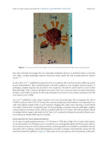
Figure 3. A T1 tumor excised with clear margins fixed on the board after transanal minimally invasive surgery excision
have been clinically downstaged but not responded completely. However it should be kept in mind that
even with a complete pathologic response of primary tumor (ypT0), the risk of nodal positivity remains
3%-6% [34-36] .
[37]
In 2016, Shin et al. published retrospective data of 34 patients who had local excision following neoad-
juvant chemoradiation. They included patients with only complete or near complete clinical response. A
pathologic complete response was achieved in 56% of patients, 35% had T1 and 9% had T2 tumor in their
final pathology. Only 2 patients developed recurrence (one local recurrence and one distant metastasis)
during a 5-year follow-up period. In this study, all lesions were located in low rectum; 28 patients had TAE
and 6 patients had TAMIS.
[30]
Lee et al. published a wider range of patients with more advanced stage. They investigated the role of
TAMIS for patients with T2/T3 N+ disease who received neoadjuvant chemoradiation and responded clini-
cally with negative lymph nodes on post treatment imaging and a final tumor showing a small whitish
scar and/or shallow ulcer on sigmoidoscopy. On final pathology, 18 patients showed a pathologic complete
clinical response of primary tumor, whereas 11 patients still had T2/T3 tumor. All of these patients refused
to undergo further surgery and nearly half of them (5 of 11 patients) developed local and/or distant recur-
rence during the median follow-up of 36 months.
Local excision for more advanced tumors
As of today, the gold standard treatment of T2-T4 lesions is TME due to high risk of lymph node metasta-
sis. Local excision of T2-T4 lesions can be considered as an oncologically inferior but less invasive alterna-
tive to radical excision in several clinical scenarios; including patients with multiple comorbidities who are
medically unfit to undergo a major abdominopelvic procedure or patients with metastatic disease but who
need local control for palliation [Figure 4]. There may also be some patients who demonstrate understand-