
Page 81 - Read Online
P. 81
Page 4 of 10 Erkan et al. Mini-invasive Surg 2018;2:30 I http://dx.doi.org/10.20517/2574-1225.2018.51
A B C
Figure 1. A T1 SM1 tumor excised by transanal minimally invasive surgery. A: Marking of resection margins with electrocautery; B:
completed full thickness excision; C: closure of rectal wall defect
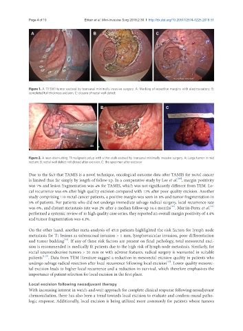
A B C
Figure 2. A near-obstructing T1 malignant polyp with a thin stalk excised by transanal minimally invasive surgery. A: Large tumor in mid
rectum; B: rectal wall defect not closed after excision; C: the specimen after excision
Due to the fact that TAMIS is a novel technique, oncological outcome data after TAMIS for rectal cancer
[29]
is limited thus far simply by length of follow-up. In a comparative study by Lee et al. , margin positivity
was 7% and lesion fragmentation was 4% for TAMIS, which was not significantly different from TEM. Lo-
cal recurrence was 6% after high quality excision compared with 13% after poor quality excision. Another
study comprising 110 rectal cancer patients, a positive margin was seen in 8% and tumor fragmentation in
5% of patients. For patients who did not undergo immediate salvage radical surgery, local recurrence rate
[30]
[31]
was 6%, and distant metastasis rate was 2% after a median follow-up 14.4 months . Martin-Perez et al.
performed a systemic review of 16 high quality case series, they reported an overall margin positivity of 4.4%
and tumor fragmentation was 4.1%.
On the other hand, another meta-analysis of 4510 patients highlighted the risk factors for lymph node
metastasis for T1 lesions as submucosal invasion > 1 mm, lymphovascular invasion, poor differentiation
[32]
and tumor budding . If any of these risk factors are present on final pathology, total mesorectal exci-
sion is recommended in medically fit patients due to the high risk of lymph node metastasis. Similarly, for
rectal neuroendocrine tumors > 20 mm or with adverse features, radical surgery is warranted in suitable
patients [1,15] . Data from TEM literature suggest a reduction in mesorectal excision quality in patients who
[33]
undergo salvage radical resection after local recurrence following local excision . Lower quality mesorec-
tal excision leads to higher local recurrence and a reduction in survival, which therefore emphasizes the
importance of patient selection for local excision in the first place.
Local excision following neoadjuvant therapy
With increasing interest in watch-and-wait approach for complete clinical response following neoadjuvant
chemoradiation, there has also been a trend towards local excision to evaluate and confirm mural patho-
logic response. Additionally, local excision is being utilized more commonly for patients whose tumors