
Page 126 - Read Online
P. 126
Myint et al. Mini-invasive Surg 2018;2:34 I http://dx.doi.org/10.20517/2574-1225.2018.52 Page 5 of 8
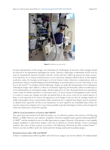
Figure 2. Treatment set up position
because interpretation of the images can sometimes be challenging. If necessary, these images should
be referred to an experienced radiologist for review. Likewise, endoscopic examination should only be
done by experienced clinicians familiar with the “watch and wait” follow-up process for these cancers.
It is important not to biopsy normal mucosa or non-cancerous radiation-induced ulcers as the negative
predictive value of a benign rectal biopsy is of very limited value. Moreover, complications such as
perforations, delays in wound healing, protracted bleeding, or persistent pain can occur if the tumor is very
[9]
low in the rectum . In addition, fibrosis following a biopsy can make the interpretation of the subsequent
radiological images more difficult. If there is uncertainty regarding abnormalities, either on endoscopy or
in the interpretation of radiological images, the best approach is to refer the patient back to an appropriate
cancer center for further assessment. In uncertain cases, it is best to repeat the investigations sooner (within
6-8 weeks) to assess any changes and refer the patient back to the cancer center for an expert opinion. If
there is local regrowth of the tumor, the appearance will change at that site, but the changes usually are
subtle and progress slowly. Examination under anesthesia for a targeted deep biopsy may be necessary
to identify local regrowth, but this is not mandatory, as most regrowth are embedded deep within the
muscles (muscularis propria) and it is not always possible to get the histological evidence of local regrowth
unless the whole area is removed surgically.
CXB for local persistence of tumors after EBCRT
The watch and wait protocol with deferred surgery can be offered to patients who achieve cCR following
EBCRT or EBRT. However, the majority of patients (74%) have residual tumor reported following EBCRT
[6]
or EBRT and the standard of care is to offer these patients surgery. However, if the patient is a not suitable
surgical candidate or still refuses surgery, CXB can be offered as a booster therapy. There is published
[4,7]
evidence that some of these patients can achieve cCR following CXB boost for their residual tumor .
Patients can then be follow up by the ‘watch and wait’ strategy and avoid immediate surgery.
Residual tumors after CXB and EBCRT
If there is residual tumor following EBRT and CXB boost, surgery can then be offered. For small residual