
Page 76 - Read Online
P. 76
Peek et al. Mini-invasive Surg 2024;8:11 https://dx.doi.org/10.20517/2574-1225.2024.47 Page 7 of 9
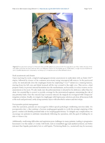
Figure 2. Port placement set-up: Dr. Sharona B. Ross and the patient are pictured with the surgical set-up of four robotic trocars: three
with eight-millimeter incisions and one with a 12-millimeter incision for the stapler device. Additionally, a single incision multi-trocar
port is used for laparoscopic instruments, involving a 2-3 cm incision and up to four different trocars.
Neck anastomosis and closure
TM
Upon reaching the neck, a stapled esophagogastrostomy anastomosis is undertaken with an Endo GIA
stapler, followed by closure of the common enterotomy using interrupted silk sutures. In the peritoneal
cavity, we meticulously close the esophageal hiatus by employing V-Loc sutures in a running fashion,
TM
starting from the left crus and distal stomach all the way around to the right crus. This serves a dual
purpose: firstly, to prevent internal herniation into the mediastinum, and secondly, to reduce tension on the
anastomosis at the neck. We also make sure the pyloromyotomy is situated in the abdomen rather than the
chest. An omental flap is created to provide coverage for the myotomized segment, minimizing the risk of
any potential leaks. Once the omental flap is securely sutured, the surgical site is irrigated with Clorpactin®
irrigation solution. Finally, we irrigate the diaphragm with bupivacaine solution and close all incisions both
at the neck and peritoneal cavity along anatomic layers with absorbable sutures and steri-strips.
Postoperative patient management
After the operation, patients are encouraged to follow speech pathologist swallowing exercises daily. On
postoperative day 3, they undergo a barium esophagogastrography to verify the prompt emptying of the
gastric conduit and identify potential leaks at the esophagogastrostomy. To mitigate any complications, we
encourage our patients to ambulate immediately following the operation, with the goal of walking for 20
min 4-5 times a day.
Additionally, swallowing difficulties and aspiration pose challenges to many patients, leading to progressive
hoarseness over the initial 4-6 weeks. Soft foods, such as scrambled eggs and mashed potatoes, are better
tolerated than liquids, particularly hot or cold liquids. Thickening fluids (i.e., nectarized) may be necessary