
Page 75 - Read Online
P. 75
Page 6 of 9 Peek et al. Mini-invasive Surg 2024;8:11 https://dx.doi.org/10.20517/2574-1225.2024.47
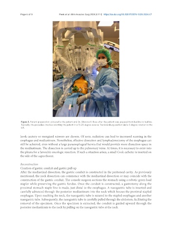
Figure 1. Patient preparation: pictured is the patient and Dr. Sharona B. Ross after the patient was prepped from bedline to bedline.
Typically, the procedure involves orienting the patient in a 15-20-degree reverse Trendelenburg position and a 5-degree rotation to the
left.
hook cautery or energized scissors are chosen. Of note, radiation can lead to increased scarring in the
esophagus and mediastinum. Nonetheless, effective dissection and lymphadenectomy of the esophagus can
still be achieved, even without a large paraesophageal hernia that would provide more dissection space in
the mediastinum. The dissection is carried up to the pulmonary veins. At times, it is necessary to enter into
the pleura for a favorable oncologic resection. If such a situation arises, a small Cook catheter is inserted on
the side of the capnothorax.
Reconstruction
Creation of gastric conduit and gastric pull-up
After the mediastinal dissection, the gastric conduit is constructed in the peritoneal cavity. As previously
mentioned, the neck dissection can commence with the mediastinal dissection or may coincide with the
construction of the gastric conduit. The console surgeon sections the stomach using a robotic green load
stapler while preserving the gastric fundus. Once the conduit is constructed, a gastrotomy along the
proximal stomach staple line is made, just distal to the esophagus. A nasogastric tube is inserted and
carefully advanced through the posterior mediastinum into the neck which houses the proximal stapled
esophagus. Upon reaching the neck, the nasogastric tube is sutured to the stapled esophagus and another
nasogastric tube. Subsequently, the nasogastric tube is carefully pulled through the abdomen, facilitating the
removal of the specimen. Once the specimen is extracted, the conduit is guided upward through the
posterior mediastinum to the neck by pulling on the nasogastric tube at the neck.