
Page 23 - Read Online
P. 23
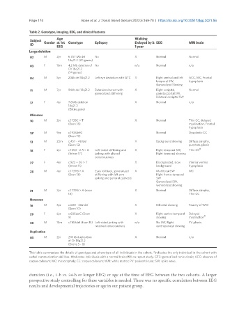
Page 174                 Bone et al. J Transl Genet Genom 2022;6:169-78  https://dx.doi.org/10.20517/jtgg.2021.56

               Table 2. Genotype, imaging, EEG, and clinical features
                            Age                                   Walking
                Subject
                ID    Gender at 1st  Genotype  Epilepsy           Delayed by ≥  EEG         MRI brain
                                                                  1 year
                            EEG
               Large deletion
                02    M     2yr   6.757 Mb del   No               X          Normal         Normal
                                  18q21.2 (20 genes)
                03    F     16m   4.2 Mb deletion of  No          n/a        Normal         n/a
                                  Ch 18q21.2
                                  (14 genes)
                04    M     5yr   20kb del 18q21.2  Left eye deviation with GTC  X  Right central and left   ACC, MC, Frontal
                                                                             temporal SW,   hypoplasia
                                                                             Generalized Slowing
                11    M     7yr   94kb del 18q21.2  Behavioral arrest with   X  Right occipital,   Normal
                                               generalized stiffening        parietoccipital SW,
                                                                             Bilateral occipital SW
                17    F     4yr   7.6Mb deletion                  X          Normal         n/a
                                  18q21.2
                                  (Entire gene)
                Missense
                10    M     2yr   c.1738C > T                     X          Normal         Thin CC, delayed
                                  (Exon 18)                                                 myelination, Frontal
                                                                                            hypoplasia
                12*   M     9yr   c.1933delG                                 Normal         Dysplastic CC
                                  (Exon 19)
                13    M     23m   c.457 - 461del                  X          Background slowing  Diffuse atrophy,
                                  (Exon 12)                                                 punctate gliosis
                                                                                                  #
                18    F     2yr   c.1650 - 2 A > G    Left-sided stiffening and   X  Right temporal SW,   Thin CC
                                  (Intron 17)  jerking with altered          Right temporal slowing
                                               consciousness
                27    F     4yr   c.922 + 3G > T                  X          Disorganized, slow   Inferior vermis
                                  (Intron 11)                                background     hypoplasia
                28    M     4yr   c.1739G > A    Eyes roll back, generalized   X  Multifocal SW   MC
                                  (Exon 18)    stiffening with left arm      Right fronto-temporal
                                               jerking and perioral cyanosis   SW
                                                                             Generalized SW,
                                                                             Generalized slowing
                31    M     3yr   c.1739G > A (exon               X          Normal         Diffuse atrophy,
                                  18)                                                       Thin CC
                Nonsense
                16    M     6yr   c.680 - 682 del                 X          Bifrontal slowing  Paucity of WM
                                  (Exon 10)
                29    F     6yr   c.605delC (Exon                 X          Right centro-temporal   Delayed
                                  9)                                         slowing        myelination #
                30    M     15m   c.1169del (Exon 15) Left-sided jerking with   n/a  No SW, Right   PV gliosis
                                               retained consciousness        centroparietal slowing
                Duplication
                05    M     2yr   201 kb duplication              X          Normal         n/a
                                  of Ch 18q21.2
                                  (Exons 5 - 8)
               This table summarizes the details of genotype and phenotype of all individuals in the cohort. *Indicates the only individual in the cohort with
               verbal communication abilities. #Indicates individuals with a normal brain MRI on repeat study. GTC: generalized tonic-clonic; ACC: absence of
               corpus callosum; MC: microcephaly; CC: corpus callosum; WM: white matter; PV: periventricular; SW: spike wave.

               duration (i.e., 1-h vs. 24-h or longer EEG) or age at the time of EEG between the two cohorts. A larger
               prospective study controlling for these variables is needed. There was no specific correlation between EEG
               results and developmental trajectories or age in our patient group.