
Page 8 - Read Online
P. 8
Fanella et al. J Transl Genet Genom 2021;5:124-9 https://dx.doi.org/10.20517/jtgg.2021.11 Page 126
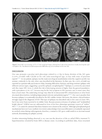
Figure 1. Brain MRI scan showing, in T2 or FLAIR-weighted images, bilateral focal subcortical alterations mostly involving the right
hemisphere (A). The alterations had disappeared at MRI follow-up performed four months later (B).
DISCUSSION
Our case presents a peculiar mild phenotype related to a 3-bp in-frame deletion of the NF1 gene
(c.2970_2972del) with CALMs as the sole extra-neurological sign, in line with some of previous
reports [7-9,11] . In our patient, epilepsy was the main neurological manifestation while the cognitive profile was
normal, contrarily to the only other case described in the literature. In that case, the c.2970_2972del was
reported in a patient with psychomotor delay, learning difficulties, dyslexia, and drug-resistant epilepsy with
[8]
different seizure types, suggesting a possible epileptic encephalopathy . Epilepsy is commonly associated
with the classic NF1 form, in which the risk of developing seizures is higher than the general population,
[6]
with a prevalence of 4%-7% . Seizures may be the first symptom in NF1 patients, and, in most cases, they
have a focal onset. The underlying etiology may often lie in intracranial NF1-related tumors or other brain
structural abnormalities, including hippocampal sclerosis and polymicrogyria [6,12,13] . Our case is peculiar first
because epilepsy is occasionally described in the NF1 milder variant. Moreover, the electro-clinical features
strongly support the diagnosis of IGE. This type of epilepsy was described only in few cases of classic NF1,
but it has never been reported in its milder form. Because presence/absence of epilepsy and “unidentified
bright objects” (UBOs) was not addressed in three of the four phenotype-genotype reports of “milder”
NF1 [7,9,10] , it cannot be assumed that these features were absent in the subjects. Although pathophysiological
mechanisms have not been clarified, it is widely acknowledged that neurofibromin is abundantly expressed
in cerebral cortex during embryogenesis; therefore, a variant in NF1 gene could result in an aberrant neural
network, determining an epileptic activity.
Another interesting finding observed in our case was the detection of the so-called UBOs, transient T2
hyperintensities revealed by brain MRI at disease onset. According to published data, these structural