
Page 107 - Read Online
P. 107
Page 244 Mohammadi et al. J Transl Genet Genom 2020;4:238-50 I https://doi.org/10.20517/jtgg.2020.29
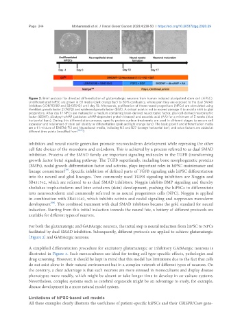
Figure 2. Brief protocol for directed differentiation of glutamatergic neurons from human induced pluripotent stem cell (hiPSC).
Undifferentiated hiPSC are grown in E8 media (dark orange bar) to 80% confluency, whereupon they are exposed to the dual SMAD
inhibitors (LDN193189 and SB431542) until day 10. Afterwards, proliferation of these neural progenitors (NPCs) are stimulated using
fibroblast growth factor 2 (FGF2) and epidermal growth factor (EGF). A critical point is not to exceed passage 4 to avoid a shift to glial
progenitors. After day 17 NPCs are matured in a medium containing brain-derived neurotrophic factor, glial cell-derived neurotrophic
factor (GDNF), dibutyryl-cAMP (activates cAMP-dependent protein kinases) and ascorbic acid (AA) for a minimum of 5 weeks (blue
horizontal bars). During this differentiation process, specific protein surface treatments are used in different stages to ensure cell
expansion and retainment of stem cell identity or differentiation (pink and light orange bars). The basic growth and differentiation media
are a 1:1 mixture of DMEM/F12 and Neuralbasal media, including N2 and B27 (orange horizontal bar), and extra factors are added at
different time points (modified from [78,79] )
inhibitors and neural rosette generation promote neuroectoderm development while repressing the other
cell fate choices of the mesoderm and endoderm. This is achieved by a process referred to as dual SMAD
inhibition. Proteins of the SMAD family are important signaling molecules in the TGFB (transforming
growth factor beta) signaling pathway. The TGFB superfamily, including bone morphogenetic proteins
(BMPs), nodal growth differentiation factor and activins, plays important roles in hiPSC maintenance and
[65]
lineage commitment . Specific inhibition of defined parts of TGFB signaling aids hiPSC differentiation
into the neural and glial lineages. Two commonly used TGFB signaling inhibitors are Noggin and
SB431542, which are referred to as dual SMAD inhibitors. Noggin inhibits BMP signaling and thereby
abolishes trophectoderm and later ectoderm (skin) development, pushing the hiPSCs to differentiate
into neuroectoderm and commonly referred to as neural progenitors cells (NPC). Noggin is applied
in combination with SB431542, which inhibits activin and nodal signaling and suppresses mesoderm
[64]
development . This combined treatment with dual SMAD inhibitors became the gold standard for neural
induction. Starting from this initial induction towards the neural fate, a battery of different protocols are
available for different types of neurons.
For both the glutamatergic and GABAergic neurons, the initial step is neural induction from hiPSC to NPCs
facilitated by dual SMAD inhibition. Subsequently, different protocols are applied to achieve glutamatergic
[Figure 2] and GABAergic neurons.
A simplified differentiation procedure for excitatory glutamatergic or inhibitory GABAergic neurons is
illustrated in Figure 3. Such monocultures are ideal for testing cell type-specific effects, pathologies and
drug screening. However, it should be kept in mind that this model has limitations due to the fact that cells
do not exist alone in their natural environment but in a complex network of different types of neurons. On
the contrary, a clear advantage is that such neurons are more stressed in monocultures and display disease
phenotypes more readily, which might be absent or take longer time to develop in co-culture systems.
Nevertheless, complex systems such as cerebral organoids might be an advantage to study, for example,
disease development in a more natural model system.
Limitations of hiPSC-based cell models
All these examples clearly illustrate the usefulness of patient-specific hiPSCs and their CRISPR/Cas9 gene-