
Page 36 - Read Online
P. 36
Chandramohan et al. J Transl Genet Genom 2024;8:394-404 https://dx.doi.org/10.20517/jtgg.2024.38 Page 400
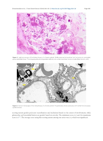
Figure 2. Light microscopy of the kidney biopsy of a female patient. Well-preserved intracellular lipid inclusions in osmicated,
epoxy-embedded tissue. The enlarged podocytes and parietal epithelial cells contain lamellated inclusion bodies (BasicFuchsin; x40).
Figure 3. Electron microscopy of the kidney biopsy of a female patient. The glomerulus shows podocytes with normal foot processes
(yellow arrows).
scoring system grades podocyte vacuolization and inclusions based on the extent of involvement, while
glomerular and interstitial lesions are graded based on severity. The minimum score is 0 and the maximum
score is 3 . The average score using this scoring system among our series was 2.4, which was significant.
[13]