
Page 76 - Read Online
P. 76
Fonseka et al. J Cancer Metastasis Treat 2020;6:7 I http://dx.doi.org/10.20517/2394-4722.2019.024 Page 5 of 11
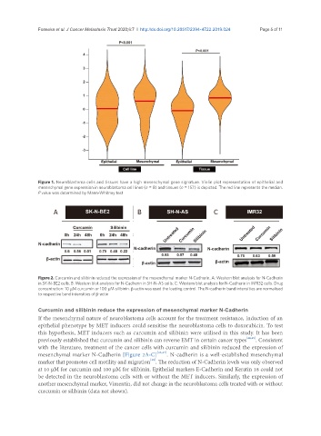
Figure 1. Neuroblastoma cells and tissues have a high mesenchymal gene signature. Violin plot representation of epithelial and
mesenchymal gene expression in neuroblastoma cell lines (n = 8) and tissues (n = 157) is depicted. The red line represents the median.
P value was determined by Mann-Whitney test
A B C
Figure 2. Curcumin and silibinin reduced the expression of the mesenchymal marker N-Cadherin. A: Western blot analysis for N-Cadherin
in SK-N-BE2 cells; B: Western blot analysis for N-Cadherin in SH-N-AS cells; C: Western blot analysis for N-Cadherin in IMR32 cells. Drug
concentration: 10 μM curcumin or 100 μM silibinin. β-actin was used the loading control. The N-cadherin band intensities are normalised
to respective band intensities of β-actin
Curcumin and silibinin reduce the expression of mesenchymal marker N-Cadherin
If the mesenchymal nature of neuroblastoma cells account for the treatment resistance, induction of an
epithelial phenotype by MET inducers could sensitise the neuroblastoma cells to doxorubicin. To test
this hypothesis, MET inducers such as curcumin and silibinin were utilised in this study. It has been
previously established that curcumin and silibinin can reverse EMT in certain cancer types [44,45] . Consistent
with the literature, treatment of the cancer cells with curcumin and silibinin reduced the expression of
mesenchymal marker N-Cadherin [Figure 2A-C] [46,47] . N-cadherin is a well-established mesenchymal
[48]
marker that promotes cell motility and migration . The reduction of N-Cadherin levels was only observed
at 10 µM for curcumin and 100 µM for silibinin. Epithelial markers E-Cadherin and Keratin 18 could not
be detected in the neuroblastoma cells with or without the MET inducers. Similarly, the expression of
another mesenchymal marker, Vimentin, did not change in the neuroblastoma cells treated with or without
curcumin or silibinin (data not shown).