
Page 466 - Read Online
P. 466
Page 18 of 40 Maner et al. J Cancer Metastasis Treat 2020;6:37 I http://dx.doi.org/10.20517/2394-4722.2020.60
The use of a TNF inhibitor, specifically etanercept, is believed to cause immune modification that may
ultimately lead to the development of cutaneous VC in a patient [119] . Patients undergoing TNF-alpha
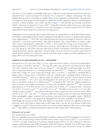
inhibitor therapy may be susceptible to complex effects on the regulation of inflammation, autoimmunity,
and apoptosis. These drugs have the potential to inhibit the extrinsic apoptotic pathway by modulating the
activation of death receptors 4 and 5 (DR4 and DR5) [Figure 7]. DR4 and DR5 are normally activated by
TRAIL, resulting in the apoptosis of cancer cells [119] . Drugs like etanercept inhibit this process, potentiating
carcinogenesis. Nonetheless, a systematic review and metanalysis of patients treated with anti-TNF-α
agents failed to demonstrate any increased risk of malignancy [120] .
Furthermore, there is a possible effect of anti-TNF therapy on hypermethylation of the ASC/TMS1 protein.
ASC/TMS1 is responsible for the activation of phagocytes through the secretion of cytokines and activation
of the inflammasome [119] . ASC/TMS1 also indirectly potentiates apoptosis through its interaction with p53
and the Bcl-2-associated X protein (BAX). BAX induces apoptosis, due to the activation of caspase 8. ASC/
TMS1 may be responsible for translocation of BAX to mitochondria by serving as its carrier protein [119] .
Hypermethylation of ASC/TMS1 inhibits these processes, enhancing cancer development. The effect of
anti-TNF drugs on ASC/TMS1 function still requires further investigation. With these known genetic
signaling pathways, pharmacogenomic treatment plans can be focused on the patient and/or lesions
specific genetic pathways in conjunction with the anti-TNF drugs to target all mutated signaling that can
appear within patients’ varying flux states.
GENETICS OF BASOSQUAMOUS CELL CARCINOMA
Basosquamous cell carcinoma (BSC) is a rare, aggressive skin neoplasm that has histopathological
characteristics of both BCC and SCC [121] . The majority of BSC cases are found in the head and neck region,
with older Caucasian males most commonly affected [122] . Clinically, BSC is indistinguishable from BCC
and is commonly referred to as “metatypical basal cell carcinoma [122,123] ”. Diagnosis of BSC is typically
made with a biopsy of a lesion suspected of being a BCC or an SCC [124] . It should be noted that BSC is
distinct from a collision tumor in that BSC is more of a “mixed tumor” consisting of BCC with areas of
SCC differentiation [125] , whereas a collision tumor has distinct BCC and SCC entities that are present
in close proximity [126] . This can be seen histologically by the presence of a transition zone of atypia in
BSC, an uncommon feature for a collision tumor [125] . Compared to BCC, BSC has a higher tendency for
local recurrence and a higher propensity for lymph node and distant metastases [121,127] . BSC comprises
approximately 2% of all skin cancers with a metastasis rate of about 7% [128] compared with a metastasis
rate of BCC of 0.0028%-0.55% [127,129] and of SCC of 2%-5% [130] . The BSC recurrence rate is 12%-51% after
standard surgical excision [128] . This contrasts with post-standard surgical excision recurrence rates of BCC
and SCC at 5%-14% [131] and ~8%, [132,133] respectively. BSC has an approximately 4% recurrence rate following
Mohs micrographic surgery compared with recurrence rates post-Mohs micrographic surgery (MMS) of
BCC (1%-2%) [131] and SCC (3%), respectively, although there was some variability in the literature [128,131,134] .
Interestingly, the recurrence rates of BSC post-MMS were reported to be about 4% regardless of site (head
and neck, trunk, lower limb, or upper limb). In BSC larger than 2 cm, the rate of recurrence is believed to
be slightly increased [128] ; however, these results were not statistically significant, likely due to small sample
size [134] . Thus, MMS is currently the preferred treatment for BSC independent of body location.
Malignant cell growth
BCC genetic drivers’ role in BSC
BSCs are frequently associated with SHH pathway mutations, implicating SHH deregulation as the primary
driver in BSC and providing evidence that BSCs shares similar cancer drivers to BCC [135] . Loss of function
in PTCH1 and gain of function in G-protein-coupled receptor SMO in the BCC pathway are the most
common mutations that cause SHH deregulation in the BCC pathway. Similarly, 45% of BSCs were shown
to have deleterious mutations in PTCH1 compared to 44% of BCCs and 10% of SCCs. About 5% of BSCs