
Page 798 - Read Online
P. 798
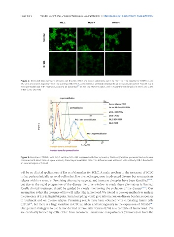
Page 4 of 6             Vander Borght et al. J Cancer Metastasis Treat 2019;5:57  I  http://dx.doi.org/10.20517/2394-4722.2019.0010


               Figure 2. Immunohistochemistry of SCLC cell line NCI-H82 and colon carcinoma cell line HCT116. The results for MUM-4 and
               MUM-6 are shown, together with the staining with RNL-1, a monoclonal antibody directed to an extracellular part of NCAM. Cells
                                                      [5]
               were permeabilized with methanol/acetone as described  or, for the MUM-6 panel, with 4% paraformaldehyde (15 min) and 0.5%
               Triton-X100 (10 min)


                            MUM-1


               Figure 3. Reaction of MUM-1 with SCLC cell line NCI-H82 measured with flow cytometry. Methanol/acetone permeabilized cells were
               compared with intact cells. A signal was only found in permeabilized cells. This difference was not found with antibody RNL-1 directed to
               an external region of NCAM

               will be on clinical applications of E18 as a biomarker for SCLC. A main problem in the treatment of SCLC
               is that patients initially respond well to first line chemotherapy, even in advanced disease, but most patients
               relapse within 6 months. Promising alternative targeted and immune therapies have been identified [10-12] ,
               but due to the rapid progression of the disease the time window to study these alternatives is limited.
               Ideally clinical treatment should be guided by closely monitoring the evolution of the disease [12,13] . Our
               assumption is that the presence of E18 will reflect the tumor load. We intend to develop methods to analyze
               the presence of E18 in liquid biopsies. Serial sampling would give information on disease burden, responses
               to treatment and on disease relapse. Promising results have been obtained with circulating tumor cells
               (CTCs) , but there is a large variation in CTC numbers and heterogeneity in the expression of NCAM .
                     [14]
                                                                                                        [14]
               Our present strategy is to use tumor derived extracellular vesicles (EVs) as a correlate of tumor load. EVs
               are constantly formed by cells, either from endosomal membrane compartments (exosomes) or from the