
Page 60 - Read Online
P. 60
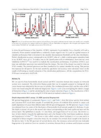
Liu et al. J Cancer Metastasis Treat 2019;5:4  I  http://dx.doi.org/10.20517/2394-4722.2018.55                                 Page 7 of 14


               Figure 3. A set of typical resonance Raman spectra collected from a horizontally sectioned normal human skin sample, and a vertically
               sliced basal cell carcinoma skin sample measured at a depth of 100 µm. Both plots are displayed in the enlarged scale regions of low
                                  -1
               wave-number 700-1,800 cm  and high wave-number 2,700-3,150 cm -1

               to show the performance of the classifier. AUROC represents the probability that a classifier will rank a
               randomly chosen positive sample before a randomly chosen negative one. It is used as a global measure of
               classifier performance that is invariant to the classifier discrimination threshold and the class distribution.
               Perfect classification accuracy corresponds to an AUROC value of 1, while a random guess separation leads
               to an AUROC value of 0.5. To reduce bias in the classification with re-substitution, leave-one-out cross
                                 [58]
               validation (LOOCV)  was used to re-evaluate the classification performance. To perform LOOCV, each
               time one individual spectrum was removed from the dataset. The rest of the dataset was used to train an
               SVM classifier. The removed spectrum was then classified by the trained classifier for testing. This process
               was repeated for all spectra. In the end, sensitivity, specificity and accuracy were calculated based on the
               results of all testing as overall evaluation of the classification performance. All the computations for PCA-
               SVM were carried out in MATLAB.


               RESULTS
               RR raw spectra from horizontally sliced normal and BCC cancerous human skin samples in vertically
               section were measured. The distinctive Raman peaks that can be uniquely assigned to distinguish skin
               cancer lesions were obtained by the raw spectral profiles. The correlation between depth and the status of BCC
               cancer was found using the RR molecular fingerprints [Figures 2 and 3], by investigating the relative changes
               of biomarkers [Figure 4] and by calculating the ratios of peak intensities [Figure 5]. The classification of BCC
               cancer from normal skin tissues using PCA-SVM statistical method is shown in Figure 6.


               Depth-dependent BCC assay: (1) VRR spectral fingerprints
               RR spectral fingerprints of carotenoids: Figures 2 and 3 and Table 1 show the typical spectra from normal
               and cancerous BCC sliced skin samples. It revealed the process of evolution from normal to cancer and
               depth-dependence of cancer from different status with RR spectral fingerprints. The RR spectrum [Figure 2 (top)]
               was obtained from the center of the third piece of horizontally sliced normal sample with thickness 50 µm.
               The depth is around 150 µm in the normal skin tissue (corresponding to the lower epidermis layer and the
               dermis layer). The epithelium is usually ~0.1 mm thick, and ranges from 0.07 to 0.12 mm. The dermis is a
               layer of skin which is beneath the epidermis layer and is the thickest of the three layers (epidermis, dermis
               and hypodermis) of skin [59-62] . The dermis is also called corium, whose thickness is 0.3-4.0 mm and it is
               composed of dense irregular connective tissue. So, we consider this normal sliced sample to be located at
               the dermis layer. In the RR spectrum of normal dermis skin, the resonance-enhanced intrinsic molecular
               fingerprints of β-carotenes (here we consider β-carotenes, because the β-carotenes and lycopene account