
Page 27 - Read Online
P. 27
Page 2 of 5 De Alcantara Filho et al. J Cancer Metastasis Treat 2019;5:2 I http://dx.doi.org/10.20517/2394-4722.2018.62
A B
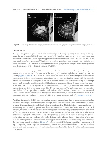
Figure 1. A: Tumor magnetic resonance imaging, axial; B: intramammary sentinel lymph node magnetic resonance imaging, axial view
CASE REPORT
A 44-year-old, post-menopausal female with a mammogram showing a partially defined lump of the right
breast. Breast ultrasound (US) showed a circumscribed hypoechoic lesion, 1.4 × 1.1 cm, 3.8 cm away from
the areolar complex, and also an adjacent hypoechoic microlobulated lesion of 1.4 × 1.2 × 1.4 cm, both in the
outer quadrants of the right breast. US-guided core-needle biopsy of the lesion revealed a high grade invasive
ductal carcinoma (IDC), luminal B (estrogen receptor 30%, progesterone receptor and human epidermal
growth factor receptor type 2 negative, and Ki-67 of 80%).
Magnetic resonance imaging (MRI) showed a mass with spiculated contours at early and heterogeneous
post-contrast enhancement in the junction of the outer quadrants of the right breast, measuring 1.8 × 1.6 ×
1.5 cm [Figure 1A and B]. In addition, a circumscribed oval mass at early and homogeneous post-contrast
enhancement, in lower inner quadrant, measuring 9 × 5 × 5 mm and in close contact with the pectoralis
muscle, which seemed to correspond to an IMLN. Second-look US was performed directly to this lymph
node, which revealed a suspicious cortical thickening and a decreased hilum. A breast conserving surgery
of the index lesion, plus radioguided occult lesion localization of the suspicious lesion in the lower inner
quadrant and sentinel lymph node biopsy (SLNB), were performed. The pathology report of the lesions
identified an IDC, no special type, histology and nuclear grade III and ductal carcinoma in situ associated.
Three axillary sentinel lymph nodes (SLNs) were free of metastases, however the suspected lesion in the
lower inner quadrant resulted in 3 IMLNs, all affected by cancer metastases with ECE [Figure 2A and B].
Published literature for IMLNs does not mention capsular leakage, thus, there is no consensus for the best
treatment. Pathologists admitted margins in lymph nodes were not frozen, which did not make it feasible
to know if the margins of the additional lesions were disease free. Multidisciplinary recommendation was
mastectomy without axillary lymph node dissection (ALND) and immediate breast reconstruction with
implants, which was performed two weeks after the patient’s consent in regards to the unknown probability
of further disease in the IMLNs. Pathology report revealed no evidence of malignancy. The patient
underwent 4 cycles of anthracycline + cyclophosphamide followed by 12 cycles of taxanes and breast +
axillary, internal mammary and supraclavicullar drainage chain radiation therapy + tamoxifen. After 2 years
of follow up, the patient suddenly developed axillary pain and fistulization accompanied by fever, and sought
the emergency room immediately [Figure 3A]. US and breast MRI showed an axillary lymph node with
high T2 signaling, measuring 4.0 × 3.6 cm in deep contact with pectoralis muscle and a circumscribed oval
mass at early and homogeneous post-contrast enhancement, located in the level 2 of the axilla, measuring
1.0 × 0.9 cm.