
Page 87 - Read Online
P. 87
Singh et al. Chest wall metastasis in head and neck carcinoma
an unusual case of squamous cell carcinoma of buccal DISCUSSION
mucosa that presented with distant metastasis to the
chest wall. Distant metastases from head and neck cancer are
unusual. The commonest site is lungs, bones, and
[9]
CASE REPORT liver and usually, occur after a long latent period. The
risk of incidence of distant metastasis depends on
A 20-year-old gentleman presented with an ulcer on the age of patient, site of the primary cancer, loco-
the right cheek. The lesion was of 2-3 months duration. regional extension, tumor grade, and loco-regional
[11]
There was associated swelling on the right side of the control by primary treatment. The risk of developing
neck region. He gave a history of tobacco chewing in distant metastasis in head and neck cancer increases
the form of gutkha (a mixture of tobacco, betel nut, and with the development of regional metastasis and is
lime) since last 7-8 years. A punch biopsy from the associated with poor survival. [12] Metastases to chest
ulcer and subsequent histopathological examination wall from head and neck cancer are extremely rare,
of the biopsy sample confirmed the diagnosis of with only a few cases reported in the literature till date.
squamous cell carcinoma of buccal mucosa. The Metastasis to such an unusual sites may be due to
patient underwent wide local excision surgery of the the disruption of lymphatic system during surgery
primary lesion with right sided modified radical neck which resulted in the lymphatic dissemination of
malignant cells to the region below the clavicle.
[13]
dissection in May 2016. Surgical pathology report was Here recurrence at the pectoralis flap site due to
suggestive of a pathological staging pT N 2b M with
0
1
lymphovascular invasion and perinodal extension.
The patient was advised adjuvant post-operative
radiotherapy with concurrent chemotherapy (cisplatin
weekly at 40 mg/m ). He received 60 Gy by external
2
beam radiotherapy in 30 fractions by conventional two-
dimensional planning on a 6 MV linear accelerator with
five cycles of concurrent cisplatin. Adjuvant therapy
was concluded in August 2016. The patient was
kept on follow-up, during which he was free of any
symptoms and signs of the disease. After a disease-
free survival of 4 months, he presented with swelling
and redness over the right side of the chest wall in
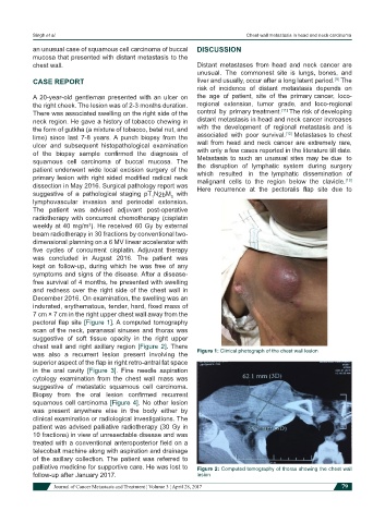
December 2016. On examination, the swelling was an
indurated, erythematous, tender, hard, fixed mass of
7 cm × 7 cm in the right upper chest wall away from the
pectoral flap site [Figure 1]. A computed tomography
scan of the neck, paranasal sinuses and thorax was
suggestive of soft tissue opacity in the right upper
chest wall and right axillary region [Figure 2]. There
was also a recurrent lesion present involving the Figure 1: Clinical photograph of the chest wall lesion
superior aspect of the flap in right retro-antral fat space
in the oral cavity [Figure 3]. Fine needle aspiration
cytology examination from the chest wall mass was
suggestive of metastatic squamous cell carcinoma.
Biopsy from the oral lesion confirmed recurrent
squamous cell carcinoma [Figure 4]. No other lesion
was present anywhere else in the body either by
clinical examination or radiological investigations. The
patient was advised palliative radiotherapy (30 Gy in
10 fractions) in view of unresectable disease and was
treated with a conventional anteroposterior field on a
telecobalt machine along with aspiration and drainage
of the axillary collection. The patient was referred to
palliative medicine for supportive care. He was lost to Figure 2: Computed tomography of thorax showing the chest wall
follow-up after January 2017. lesion
Journal of Cancer Metastasis and Treatment ¦ Volume 3 ¦ April 28, 2017 79