
Page 93 - Read Online
P. 93
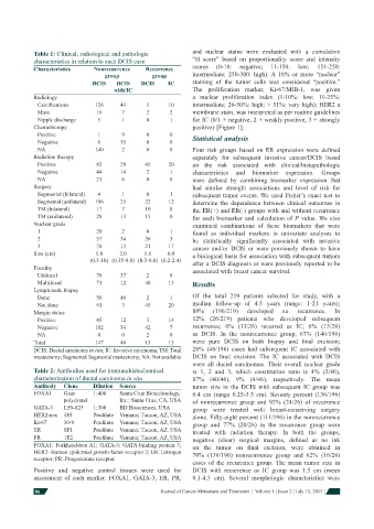
Table 1: Clinical, radiological and pathologic    and nuclear stains were evaluated with a cumulative
            characteristics in relation to each DCIS case     “H score” based on proportionality score and intensity
            Characteristics    Nonrecurrence   Recurrence     scores  (0-10:  negative;  11-150:  low;  151-250:
                                  group           group       intermediate; 250-300: high). A 10% or more “nuclear”
                              DCIS    DCIS    DCIS     IC     staining of the tumor cells was considered “positive.”
                                     with/IC                  The proliferation marker, Ki-67/MIB-1, was given
            Radiology                                         a nuclear proliferation index (1-10%: low; 11-25%:
             Calcifi cations    126     41       1      10     intermediate; 26-50%: high; > 51%: very high); HER2 a
             Mass               16      7       2      2      membrane stain, was interpreted as per routine guidelines
             Nipple discharge   5       1       0      1      for IC (0/1 + negative, 2 + weakly positive, 3 + strongly
            Chemotherapy                                      positive) [Figure 1].
             Positive           1       9       0      0      Statistical analysis
             Negative           6      35       0      0
             NA                140      2       0      0      Four risk groups based on ER expression were defi ned
            Radiation therapy                                 separately for subsequent invasive cancer/DCIS based
             Positive           82     29      45      20     on the risk associated with clinical/histopathologic
             Negative           44     14       2      1      characteristics and biomarker expression. Groups
             NA                 21      6       0      0      were defi ned by combining biomarker expression that
            Surgery                                           had similar strength associations and level of risk for
             Segmental (bilateral)  4   1       0      1      subsequent tumor events. We used Fisher’s exact test to
             Segmental (unilateral)  106  25   22      12     determine the dependence between clinical outcomes in
             TM (bilateral)     17      7      10      8      the ER(+) and ER(-) groups with and without recurrence
             TM (unilateral)    28     13      15      0      for each biomarker and calculation of P  value. We  also
            Nuclear grade                                     examined combinations of these biomarkers that were
             1                  20      2       0      1      found as individual markers in univariate analyses to
             2                  57     34      26      3      be statistically signifi cantly associated with invasive
             3                  70     13      21      17     cancer and/or DCIS or were previously shown to have
            Size (cm)          1.8     2.0     1.6    0.8     a biological basis for association with subsequent tumors
                              (0.3-10) (0.35-9.0) (0.3-9.0) (0.2-2.4)  after a DCIS diagnosis or were previously reported to be
            Focality                                          associated with breast cancer survival.
             Unifocal           76     37       2      8
             Multifocal         71     12      40      13     Results
            Lymph node biopsy
             Done               56     46       2      1      Of the total 219 patients selected for study, with a
             Not done           91      3      45      20     median follow-up of 4.5 years (range: 1-21 years);
            Margin status                                     88%    (196/219)  developed  no   recurrence.  In
             Positive           45     12       3      14     12% (26/219) patients who developed subsequent
             Negative          102     34      42      7      recurrence; 6% (13/26) recurred as IC; 6% (13/26)
             NA                 0       0       2      0      as DCIS. In the nonrecurrence group, 67% (146/196)
            Total              147     46      13      13     were pure DCIS on both biopsy and  fi nal  excision;
            DCIS: Ductal carcinoma in situ; IC: Invasive carcinoma; TM: Total   26% (46/196) cases had subsequent IC associated with
            mastectomy; Segmental: Segmental mastectomy; NA: Not available  DCIS on  fi nal excision.    The IC associated with DCIS
                                                              were all ductal carcinomas.  Their overall nuclear grade
            Table 2: Antibodies used for immunohistochemical   is 1, 2 and 3, which constitution ratio is 4% (2/46),
            characterization of ductal carcinoma in situ      87% (40/46), 9% (4/46), respectively.  The mean
            Antibody  Clone    Dilution  Source               tumor size in the DCIS with subsequent IC group was
            FOXA1    Goat      1:400   Santa Cruz Biotechnology,   0.4 cm (range 0.25-3.5 cm). Seventy percent (136/196)
                     polyclonal        Inc.; Santa Cruz, CA, USA  of nonrecurrence group and 92% (24/26) of recurrence
            GATA-3   L50-823   1:300   BD Biosciences, USA    group were treated with breast-conserving surgery
            HER2-neu  485      Predilute  Ventana; Tucson, AZ, USA  alone. Fifty-eight percent (111/196) in the nonrecurrence
            Ki-67    30-9      Predilute  Ventana; Tucson, AZ, USA  group and 77% (20/26) in the recurrence group were
            ER       SP1       Predilute  Ventana; Tucson, AZ, USA  treated with radiation therapy. In both the groups,
            PR       1E2       Predilute  Ventana; Tucson, AZ, USA  negative (clear) surgical margins, defi ned as no ink
            FOXA1: Forkhead-box A1; GATA-3: GATA binding protein 3;   on the tumor on  fi nal excision, were obtained in
            HER2: Human epidermal growth factor receptor 2; ER: Estrogen   70% (136/196) nonrecurrence group and 62% (16/26)
            receptor; PR: Progesterone receptor
                                                              cases of the recurrence group.  The mean tumor size in
            Positive and negative control tissues were used for   DCIS with recurrence as IC group was 1.5 cm (mean
            assessment of each marker. FOXA1, GATA-3, ER, PR,   0.1-4.5 cm). Several morphologic characteristics were

            86                                      Journal of Cancer Metastasis and Treatment  ¦  Volume 1 ¦ Issue 2 ¦ July 15, 2015 ¦