
Page 76 - Read Online
P. 76
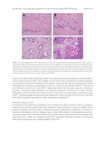
Skorupan et al. J Cancer Metastasis Treat 2023;9:5  https://dx.doi.org/10.20517/2394-4722.2022.106  Page 5 of 26


                Figure 1. Histologic appearance of ASCP. H&E specimens of ASCP. (A) Areas resembling conventional ductal adenocarcinoma of the
                pancreas (top right) and squamous cell carcinoma (bottom left) are apparent in this sample (100×). Note the striking amount of
                desmoplastic stroma present between tumor nests. (B) In this area of the tumor, the glandular and squamous components have
                merged together so that both morphologies can be seen within a single group of tumor cells (100×). (C) Infiltrative glands making up
                the adenocarcinoma component of the tumor are shown at higher power (200×), with some cells containing possible mucin vacuoles.
                (D) Solid sheets of tumor cells with densely eosinophilic cytoplasm and areas of keratinization, consistent with the squamous cell
                carcinoma component of the tumor, are shown at higher power (200×).


               to giant cell or pleomorphic-appearing anaplastic carcinomas with multi-nucleated forms, markedly bizarre
                                          [45]
               nuclei, and prominent nucleoli . Interestingly, a recent study of the transitional area between glandular
               and squamous components identified an intermediate component that appeared glandular but stained
                                                                                             [55]
               heterogeneously for squamous and glandular markers (i.e., CK8/18 and p63 double-positive) . This type of
               dual staining was also seen in some PDAC samples that lacked any histologic squamous component.
               Recently, a retrospective study identified a sarcomatous component to ASCP in 7 of 7 cases , but this
                                                                                                [54]
               finding has not been mentioned by others except for one case report describing a particularly large
               ASCP . Immunologically, the adenocarcinoma component of ASCP resembles PDAC; however, ASCP has
                    [56]
               been noted to frequently express PD-L1 within the squamous component [57,58] .


               Molecular profiling of ASCP
               Normal pancreatic tissue has no squamous cells, leading to the obvious question of how a squamous
               component can appear in a primary pancreatic malignancy. Three etiologies were proposed: collision of two
               separate primaries to form one tumor, metaplasia caused by inflammation resulting in a transition to
               squamous differentiation in the pre-malignant stage, or trans-differentiation of existing adenocarcinoma to
               a squamous component . Presently, the latter theory is favored given the genomic and transcriptomic data
                                   [59]
               described  below,  which  are  consistent  with  ASCP  appearing  following  activation  of  squamous
               differentiation programs in a pre-existing epithelial cancer cell [35,39] .