
Page 45 - Read Online
P. 45
Balakrishnan et al. J Cancer Metastasis Treat 2022;8:27 https://dx.doi.org/10.20517/2394-4722.2022.33 Page 5 of 17
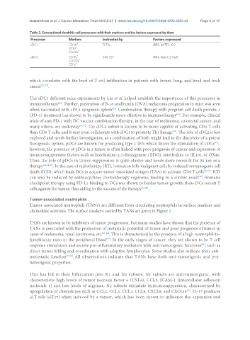
Table 2. Conventional dendritic cell precursors with their markers and the factors expressed by them
Precursor Markers Instructed by Factors expressed
cDC1 CD141 + FLT3L IRF8, BATF3, ID2
+
XCR1
+
Clec9a
+
cDC2 CD11b GM-CSF IRF4, Notch 2, RelB
+
CD172a
CD1c +
which correlates with the level of T cell infiltration in patients with breast, lung, and head and neck
cancer [25-27] .
The cDC1 deficient mice experiments by Liu et al. helped establish the importance of this precursor in
immunotherapy . Further, prevention of B-16 ovalbumin (OVA) melanoma progression in mice was seen
[28]
[29]
when vaccinated with cDC1 syngeneic spleen . Combination therapy with program cell death protein-1
(PD-1) treatment has shown to be significantly more effective in immunotherapy . For example, clinical
[20]
trials of anti-PD-1 with DC vaccine combination therapy, in the case of melanoma, colorectal cancer, and
many others, are underway [30,31] . The cDC2 subset is known to be more capable of activating CD4 T cells
than CD8 T cells, and it may even collaborate with cDC1 to promote Th1 lineage . The role of cDC2 is less
[32]
explored and needs further investigation, as a combination of both might lead to the discovery of a potent
therapeutic option. pDCs are known for producing type 1 IFN which drives the stimulation of cDCs ;
[33]
however, the presence of pDCs in a tumor is often linked with poor prognosis of cancer and expression of
immunosuppressive factors such as Indolamine-2,3-dioxygenase 1(IDO), interleukin-10 (IL10), or OX40.
Thus, the role of pDCs in tumor suppression is quite elusive and needs more research for its use as a
therapy [32,34,35] . In the case of radiotherapy (RT), ionization kills malignant cells by induced immunogenic cell
death (ICD), which leads DCs to acquire tumor-associated antigen (TAA) to activate CD8 T cells [36,37] . ICD
+
can also be induced by anthracyclines chemotherapy regimens, leading to a similar result . Immune
[38]
checkpoint therapy using PD-L1 binding to DCs was shown to hinder tumor growth; these DCs recruit T
cells against the tumor, thus aiding in the success of the therapy [39,40] .
Tumor-associated neutrophils
Tumor-associated neutrophils (TANs) are different from circulating neutrophils in surface markers and
chemokine activities. The surface markers carried by TANs are given in Figure 1.
TANs are known to be inhibitors of tumor progression, but many studies have shown that the presence of
TANs is associated with the promotion of metastatic potential of tumor and poor prognosis of tumor in
cases of melanoma, renal carcinoma, etc. [41-44] . This is characterized by the presence of a high neutrophil-to-
[45]
lymphocyte ratio in the peripheral blood . In the early stages of cancer, they are shown to be T cell
response stimulators and secrete pro-inflammatory mediators with anti-tumorigenic functions , such as
[46]
direct tumor killing and coordination with adaptive lymphocytes. Some studies also indicate their anti-
metastatic function [47,48] . All observations indicate that TANs have both anti-tumorigenic and pro-
tumorigenic properties.
This has led to their bifurcation into N1 and N2 subsets. N1 subsets are anti-tumorigenic, with
characteristic high levels of tumor necrosis factor α (TNFα), CCL3, ICAM-1 (intercellular adhesion
molecule 1) and low levels of arginase. N2 subsets stimulate immunosuppression, characterized by
upregulation of chemokines such as CCL2, CCL3, CCL4, CCL8, CXCL8, and CXCL16 . IL-17 produces
[49]
γδ T cells (γδT17) when induced by a tumor, which has been shown to influence the expansion and