
Page 29 - Read Online
P. 29
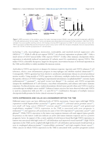
Rabadi et al. J Cancer Metastasis Treat 2022;8:24  https://dx.doi.org/10.20517/2394-4722.2022.06  Page 3 of 14


                Figure 1. VISTA expression in the periphery versus the tumor microenvironment. BALB/c mice were injected intradermally with 100k
                CT26 cells and were grown for two weeks. Tumors and spleens were dissociated and processed for flow cytometry. Cells from VISTA-
                deficient mice were used for staining controls. (A) Overlays showing VISTA expression on CD8+ T cells in the spleen or tumor. (B)
                VISTA expression as Mean Fluorescence intensity in spleen versus tumor within the indicated cell subsets. Data are represented as
                mean ± SD. VISTA: V-domain Ig Suppressor of T cell Activation.

               including  T  cells,  macrophages,  monocytes,  neutrophils,  and  myeloid-derived  suppressor  cells
                                                                                                 [21]
                                                            [19]
               (MDSCs) [15,19,20] . While B cells do not express VISTA , we observed expression on plasma cells . Only a
               small subset of CD56 natural killer (NK) express VISTA . Unlike other immune checkpoints whose
                                                                  [19]
                                  hi
               expression is selectively induced on particular IC subsets, most ICs constitutively express VISTA. This
               makes VISTA a desirable therapeutic target for therapeutic intervention because of its broad expression in
               the TME and its ability to widely modulate the immune system.
               Antibodies to VISTA can improve or dampen the immune response. Agonistic anti-VISTA enhances T cell
               tolerance, elicits a non-inflammatory program in macrophages and inhibits myeloid chemotaxis [12,22] .
               Consequently, VISTA agonism has been shown to ameliorate autoimmune disease in several preclinical
               murine models. Using models of VISTA agonism or deficiency, multiple studies have demonstrated the
               therapeutic relevance of VISTA targeting in experimental autoimmune encephalomyelitis , systemic lupus
                                                                                           [23]
               erythematosus [23-25] , psoriasis  , and graft versus host disease [13,27] . In contrast, anti-VISTA antagonists
                                       [26]
               dampen the establishment of T cell tolerance, exacerbate inflammation, and enhance anti-tumor immune
               responses. Preclinical murine cancer studies demonstrate slowed tumor growth when anti-VISTA is used as
                                                  [9]
               a monotherapy in multiple cancer models . Enhanced tumor rejection has been observed when anti-VISTA
               is used in conjunction with anti-PD-1 or anti-PD-L1 . Combination therapies of multiple immune
                                                               [28]
               checkpoints holds promise for better clinical outcomes for patients.

               VISTA EXPRESSION AND VALUE AS A BIOMARKER WITHIN THE TME
               Different tumor types can have differing levels of VISTA expression. Cancer types with high VISTA
               expression include hepatocellular carcinoma [29,30] , gastric cancers [31,32] , colorectal cancer, prostate cancer ,
                                                                                                       [11]
                                                                                        [35]
               non-small  cell  lung  cancer  (NSCLC) , ovarian  cancer , endometrial  cancer , and  gestational
                                                                   [34]
                                                   [33]
               trophoblastic  neoplasia . VISTA  expression  in  the  TME  is  associated  with  poor  prognosis  in
                                     [36]
                                                                                  [39]
               melanoma [37,38] , oral squamous cell carcinoma (in cases of low CD8 expression) , pancreatic cancer , and
                                                                                                    [40]
               gliomas . While VISTA expression in the tumor may suppress immune cells and support tumor growth,
                      [41]
               its presence on the tumor could also indicate an active anti-tumor immune response and be a positive
               prognostic factor. In support of this, a meta-analysis of solid tumors found that high VISTA expression in
               tumors was associated with high levels of T cell infiltration and improved overall survival . High VISTA
                                                                                            [42]
               expression in the TME can be coupled with high levels of T cell infiltration and increased overall survival, as
               seen in esophageal adenocarcinoma , hepatocellular carcinoma  and triple-negative breast cancer .
                                                                       [30]
                                                                                                       [44]
                                              [43]
               Other cancers in which VISTA expression in the TME is associated with a positive prognosis include