
Page 69 - Read Online
P. 69
Page 6 of 9 Shaha. J Cancer Metastasis Treat 2023;9:22 https://dx.doi.org/10.20517/2394-4722.2022.101
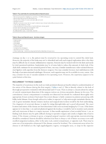
Table 5. Tips and tricks for neck dissection in thyroid cancer
• Review preoperative imaging very carefully - CT/MRI/Ultrasound
• Review thyroid bed and paratracheal area
• Preoperative status of vocal cords and calcium levels
• Necklace incision
• Identify accessory nerve, but no need to skeletonize or dissect above the accessory nerve
• Look for jugulodigastric nodes
• Avoid dissection on the surface of submandibular salivary gland
Look for supraclavicular and retrojugular node
Look for pre- and paratracheal nodes
Avoid lymphatic injury - chyle leak, chyloma
Table 6. Recurrent thyroid cancer - decision octagon
• Undetectable
• Detectable
• Actionable
• Prognosticating
• Critical location
• Complications of surgery
• Opportune time for surgery
• Close monitoring - recurrence may lead to further detection of recurrence
drainage on day 1 or 2, the patient may be returned to the operating room to control the chyle leak.
However, the majority of the leaks may not be identified and early and surgical exploration after a few days
may be difficult due to intense inflammatory response. Patients may be treated with low-fat diets and rarely
by total parenteral nutrition. Sandostatin may be of some help to reduce the amount of chyle leak. If the
chyle leak continues for an extended period of time, one may consider transthoracic video-assisted thoracic
duct ligation with expertise from the thoracic surgeon. One may consider thoracic duct embolization with
the help of an interventional radiologist. However, such expertise may not be available in every center. One
may consider the use of vascular sealants in the operating room. However, the experience appears to be
quite limited at this time.
RECURRENT THYROID CANCER
The majority of recurrences in the neck are truly persistent diseases primarily related to underestimation of
the extent of the disease during the first surgery [Tables 6 and 7]. This is directly related to the lack of
thorough preoperative evaluation with ultrasound and CT scan. There is always a concern about the extent
of central compartment dissection if the surgeon notices an obvious metastatic disease in this area. The
contralateral central compartment is routinely not dissected but should be evaluated thoroughly with
appropriate frozen sections if indicated. Recurrent disease is fairly common in patients presenting with
bulky nodal disease. Even though radioactive iodine is expected to help microscopic metastatic disease, its
role in gross metastatic disease remains unclear and surgical intervention would be the best undertaking.
The diagnosis of recurrent disease is made by rising thyroglobulin and a good ultrasound. The exact
location and extent of the disease are best determined with cross-sectional imaging by CT scan. If the lesion
appears to be less than 1 cm and not plastered against the trachea, it would be best to monitor such patients
and only intervene if there is a major increase. A fine-needle aspiration is best avoided below 1 cm, as
finding such disease may be quite difficult during re-operative surgery and clearly at higher risk of nerve
injury. If the disease continues to grow, a targeted surgical resection with appropriate neuromonitoring
should be considered. Patients should be informed that there is always a risk of future recurrence even with
excellent surgical procedures, either in the same neck, central compartment, or opposite neck. There may be
some use of intraoperative ultrasound if the disease cannot be localized. The commonest areas where the
disease is left behind are the high jugulodigastric region, retrojugular area, or deep in the superior