
Page 68 - Read Online
P. 68
Shaha. J Cancer Metastasis Treat 2023;9:22 https://dx.doi.org/10.20517/2394-4722.2022.101 Page 5 of 9
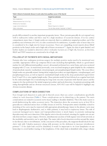
Table 4. Extent of metastatic disease in neck nodes from papillary cancer of the thyroid
Type Import on outcome
Micrometastasis None
Mini metastasis (by U/S of Tg) None
Minivolume metastasis None
Large volume metastasis Maybe (regional or distant)
Major metastasis Yes, in older patients (regional or distant)
poorly differentiated) is another important prognostic factor. These patients generally do not respond very
well to radioactive iodine and there may be a high incidence of recurrent disease. If in the central
compartment, more than 11 lymph nodes are removed, that is a satisfactory surgical procedure, and if the
positivity rate is between 20%-50%, it is also considered to be satisfactory. A positive ratio of more than 65%
is considered to be a high risk for future recurrence. There are compelling recent reports about BRAF
positivity in the lymph nodes and a high risk of future recurrences . Again, the data is quite limited, and
[20]
further studies are needed to determine which molecular markers are of high risk, however, at this time,
BRAF and TERT mutation are considered to be of high risk.
FOLLOW-UP OF PATIENTS WITH NODAL METASTASIS
Patients who have undergone previous surgery for multiple positive nodes need to be monitored very
carefully. Appropriate follow-up comprises blood tests (including thyroglobulin, which is a good tumor
marker for well-differentiated papillary cancer), ultrasound performed on a serial basis, and cross-sectional
imaging with CT scan. As mentioned previously, cross-sectional imaging is quite helpful for the evaluation
of paratracheal, superior mediastinal, retropharyngeal, and parapharyngeal lymph nodes. The areas where
lymph nodes are generally missed during initial surgical procedure are jugulodigastric, retropharyngeal, and
parapharyngeal areas, as well as superior mediastinal lymph nodes in the deep paratracheal region below
level VI and VII or retro jugular lymph nodes. These patients would be best followed on a regular basis both
by the endocrinologist who is monitoring the long-term outcome of patients with thyroid cancer and the
surgeon who has performed the initial surgical procedure and neck dissection. In patients presenting with
rising thyroglobulin and no obvious structural disease, FDG PET scan will be helpful to highlight any
obvious recurrent disease.
COMPLICATIONS OF NECK DISSECTION
Even though neck dissection is quite safe in thyroid cancer, there are certain complications specifically
related to neck dissection [Table 5]. These complications can be disturbing to the patient. The main
complications are related to accessory nerve injury leading to shoulder disability. This is the main reason to
avoid skeletonizing the entire accessory nerve. The dissection above the accessory nerve at level IIb is
generally not indicated unless there is bulky disease at level IIa. Postoperative minor disability related to
stretching of the nerve may be improved with appropriate physiotherapy. Injury to the phrenic nerve may
lead to diaphragmatic dysfunction, and in older patients, it may lead to respiratory issues. The risk of
recurrent laryngeal nerve injury and hypoparathyroidism is much higher in patients undergoing
thyroidectomy with neck dissection. If the jugular vein is directly involved, resecting the jugular vein on one
side does not have a major impact. However, simultaneous resection of both jugular veins is best avoided, as
the morbidity and mortality can be quite high. The major risk of neck dissection revolves around the injury
to the thoracic duct or its tributaries. At the conclusion of the procedure, the surgeons should check for any
chyle leak with Valsalva maneuver along with pressure on the abdomen to increase intrathoracic
pressure . If the chyle leak could be identified, it would be important to tie these lymphatics or use
[15]
hemoclips to avoid further tearing of the lymphatics. If the chyle leak is identified with a large volume