
Page 8 - Read Online
P. 8
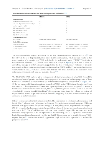
Atmaja et al. J Cancer Metastasis Treat 2021;7:xx  https://dx.doi.org/10.20517/2394-4722.2021.66  Page 3 of 14

               Table 1. Differences between the MSKCC and IMDC risk prognostication tools for mRCC [6-8]
                                              MSKCC                          IMDC
                Prognostic factors
                Low Karnofsky performance (< 80%)  √                         √
                Time from diagnosis to treatment < 1 year  √                 √
                Low haemoglobin (< LLN)       √                              √
                High corrected calcium (> ULN)  √                            √
                High neutrophils (> ULN)                                     √
                High platelets (> ULN)                                       √
                High LDH (> 1.5 times ULN)    √
                Entry population criteria
                MRCC patient                  Interferon as frontline therapy  First-line TKI therapy
                Median OS (by risk groups)
                0 criteria (favorable)        29.6 months (95%CI: 20.9-37.8 months)  43.2 months (95%CI: 31.4-50.1 months)
                1-2 criteria (intermediate)   13.8 months (95%CI: 12.4-15.9 months)  22.5 months (95%CI: 18.7-25.1 months)
                ≥ 3 criteria (poor)           4.9 months (95%CI: 4.3-6.3 months)  7.8 months (95%CI: 6.5-9.7 months)


               The inactivation of von Hippel-Lindau (VHL) is the most common mutation observed in ccRCC . The
                                                                                                    [15]
               loss of VHL leads to hypoxia-inducible factor (HIF) accumulation, which in turn results in the
               overexpression of pro-angiogenic VEGF and platelet-derived growth factor (PDGF) [15,16] . Sunitinib, a
               tyrosine kinase Inhibitors (TKI), blocks VEGF and PDGF receptors [Figure 1] ; it is used as a first or
                                                                                    [17]
               second-line therapy in ccRCC. Research suggests that the loss of VHL is not sufficient to promote
               oncogenesis, and that mutations of epigenetic regulators such as PBRM1 and BAP1 are required to drive the
                                    [18]
               development of ccRCC . Studies indicate that tumors with BAP1 mutations are correlated with
               unfavorable outcomes in both local and metastatic disease [19,20] .

               The PI3K/AKT/mTOR pathway plays an important role in the tumorigenesis of ccRCC. The mTOR
               complex regulates cell growth, metabolism and angiogenesis; mutations can lead to dysregulation of these
               functions . Everolimus and Temsirolimus are mTOR inhibitors approved for use in mRCC [Figure 1] .
                                                                                                       [22]
                       [21]
               Of the two known mTOR signaling pathways, namely mTORC1 and mTORC2, these therapies only block
               one (mTORC1), leaving the second downstream signaling of mTOR activation unopposed [23,24] . Research
               has identified that tumor mutations in mTOR, TSC1 or 2 (mTOR regulators) are more common in patients
                                                      [25]
               who clinically respond to mTOR inhibitors . However, one study found that a large proportion of
               responders had no mTOR pathway mutation detected , suggesting that these mutations cannot yet be
                                                               [25]
               considered as useful biomarkers.

               ICIs are commonly used in the treatment of mRCC. The combination of Nivolumab, a Programmed Cell
               Death (PD-1) inhibitor and Ipilimumab, a Cytotoxic T-Lymphocyte-associated Antigen 4 (CTLA-4)
               inhibitor, is an approved first-line systemic therapy . In many malignancies, Programmed Death-Ligand 1
                                                          [26]
               (PD-L1) expression has been demonstrated as a reliable predictor of therapy response; however, this is not
               the  case  in  RCC.  The  CheckMate-025  and  -214  trials  recorded  respectable  response  rates  with
               immunotherapy regardless of PD-L1 expression [27-29] . Unfortunately, its use as a potential biomarker is
               limited because of tumor heterogeneity and the fact that PD-L1 expression can be modified by prior VEGF-
               targeted therapies . Nevertheless, research suggests that a high level of PD-L1 expression is an unfavorable
                              [30]
               prognostic factor [30,31] .