
Page 25 - Read Online
P. 25
Quartuccio et al. J Cancer Metastasis Treat 2021;7:14 I http://dx.doi.org/10.20517/2394-4722.2020.118 Page 5 of 13
18
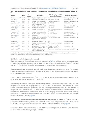
Table 1. Main characteristics of studies with patients with thyroid cancer and hematogenous metastases evaluated by F-FDG PET/
CT
No. Other imaging modalities
Authors Years Histotype Standard of reference
patients used for comparison
Freudenberg et al. [19] 2007 36 Papillary (21), Follicular (15) CT Histology, clinical follow-up
18
Klain et al. [20] 2020 40 Differentiated (papillary = 30) MRI, F-FDG PET/MRI Clinical follow-up
Kundu et al. [21] 2015 62 Papillary (56), Follicular (6) 68 Ga-DOTANOC PET/CT Histology, clinical follow-up
131
Leboulleux et al. [22] 2012 34 Papillary (32), Follicular (2), CT, I-WBS Clinical follow-up
Poorly differentiated (2)
131
Nagamachi et al. [23] 2011 70 Papillary (62), Follicular (8) MRI, I-WBS Histology, clinical follow-up
Nakajo et al. [24] 2013 20 Papillary (19), Follicular (1) 18 F-FLT PET/CT Clinical follow-up
Ota et al. [25] 2013 11 Papillary (4), Follicular (7) Bone scan (planar and SPECT), Clinical follow-up
18 F-fluoride PET/CT
Piccardo et al. [26] 2011 20 Papillary (12), Follicular (8) 131 I-WBS, C.I. Clinical follow-up
Piccardo et al. [27] 2019 25 Papillary (18), Follicular (7) 18 F-choline PET/CT Histology, clinical follow-up
Poisson et al. [28] 2010 20 Anaplastic CT Clinical follow-up
Qiu et al. [29] 2012 80 Papillary (37), Follicular (39), 131 I-SPECT/CT, bone scan Histology, clinical follow-up
Follicular variant of papillary
(4)
Sakurai et al. [30] 2012 23 Papillary (6), Follicular (17) MRI Clinical follow-up
Shinto et al. [31] 2015 28 Papillary (10), Follicular (14), 99m Tc-Hynic TOC SPECT/CT Clinical follow-up
Hurtle cell (4)
68
Vrachimis et al. [33] 2016 26 Differentiated, with suspicion 18 F-FDG PET/MRI, Ga- Clinical follow-up
or known dedifferentiation DOTANOC PET/CT
Vrachimis et al. [34] 2016 12 Papillary (5), Follicular (3) 68 Ga-DOTATATE PET/MRI, MRI Histology, clinical follow-up
Poorly differentiated (4)
131
No.: Number; CT: computed tomography; MRI: magnetic resonance imaging; I-WBS: 131-Iodium whole-body scan; C.I.: conventional
imaging; FLT: fluorothymidine; SPECT: single photon emission computed tomography.
Qualitative analysis (systematic review)
The characteristics of the 15 selected studies are presented in Table 1. All these articles were single-center
investigations, published within the last 10 years, except one (2007), by authors from Europe (n = 8) and
Asia (n = 7). Two thirds of the studies were retrospective and one third were prospective.
The patient sample was consistently relatively small across the studies ranging from 11 to 80. The histotype
most represented was papillary (67%), followed by follicular (27%). Only one study recruited exclusively
patients with anaplastic histotype.
18
In the 15 studies, patients underwent F-FDG PET/CT scan at different moments of the diagnostic work-
131
up before or after at least one cycle of I treatment.
The hematogenous lesions investigated were located prevalently in bone and lung. In each study, PET was
18
compared with at least one imaging modality. In four studies, F-FDG PET/CT was assessed in a head-
to-head comparison with MRI, prevalently with diffusion-weighted imaging (DWI). CT was available for
18
comparison with F-FDG PET/CT in three studies. Patients underwent both PET/MRI and PET/CT in
three studies. The standard of reference was based on clinical imaging follow-up in most studies; histology
was adopted as additional standard of reference in six studies. The risk of bias for the studies was scored as
low by using the QUADAS-2 [Figure 2].
18
Meta-analysis: detectability of hematogenous metastatic lesions by means of F-FDG PET/CT
18
Considering the five studies (patients = 84) for which patient-based analysis was available, F-FDG PET/
CT detected hematogenous metastases in 85.08% (95% C.I.: 75.96%-91.75%) of cases [Figure 3].
18
Considering the 691 lesions (14 studies), F-FDG PET/CT showed a pooled DR of 89.70% (95%CI: 79.61%-
2
96.62%) with substantial heterogeneity (I = 92.84%).