
Page 64 - Read Online
P. 64
Page 8 of 16 Karolak et al. J Cancer Metastasis Treat 2021;7:15 https://dx.doi.org/10.20517/2394-4722.2021.05
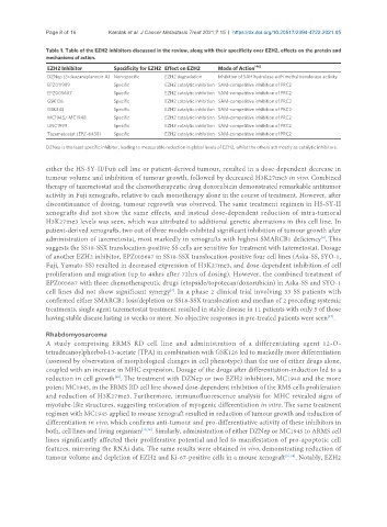
Table 1. Table of the EZH2 inhibitors discussed in the review, along with their specificity over EZH2, effects on the protein and
mechanisms of action.
EZH2 Inhibitor Specificity for EZH2 Effect on EZH2 Mode of Action [46]
DZNep (3-deazaneplanocin A) Non-specific EZH2 degradation Inhibition of SAH hydrolase with methyltransferase activity
EPZ011989 Specific EZH2 catalytic inhibition SAM-competitive inhibition of PRC2
EPZ005687 Specific EZH2 catalytic inhibition SAM-competitive inhibition of PRC2
GSK126 Specific EZH2 catalytic inhibition SAM-competitive inhibition of PRC2
GSK343 Specific EZH2 catalytic inhibition SAM-competitive inhibition of PRC2
MC1945/ MC1948 Specific EZH2 catalytic inhibition SAM-competitive inhibition of PRC2
UNC1999 Specific EZH2 catalytic inhibition SAM-competitive inhibition of PRC2
Tazemetostat (EPZ-6438) Specific EZH2 catalytic inhibition SAM-competitive inhibition of PRC2
DZNep is the least specific inhibitor, leading to measurable reduction in global levels of EZH2, whilst the others act mostly as catalytic inhibitors.
either the HS-SY-II/Fuji cell line or patient-derived tumour, resulted in a dose-dependent decrease in
tumour volume and inhibition of tumour growth, followed by decreased H3K27me3 in vivo. Combined
therapy of tazemetostat and the chemotherapeutic drug doxorubicin demonstrated remarkable antitumor
activity in Fuji xenografts, relative to each monotherapy alone in the course of treatment. However, after
discontinuance of dosing, tumour regrowth was observed. The same treatment regimen in HS-SY-II
xenografts did not show the same effects, and instead dose-dependent reduction of intra-tumoral
H3K27me3 levels was seen, which was attributed to additional genetic aberrations in this cell line. In
patient-derived xenografts, two out of three models exhibited significant inhibition of tumour growth after
administration of tazemetostat, most markedly in xenografts with highest SMARCB1 deficiency . This
[4]
suggests the SS18-SSX translocation-positive SS cells are sensitive for treatment with tazemetostat. Dosage
of another EZH2 inhibitor, EPZ005687 in SS18-SSX translocation-positive four cell lines (Aska-SS, SYO-1,
Fuji, Yamato-SS) resulted in decreased expression of H3K27me3, and dose-dependent inhibition of cell
proliferation and migration (up to 48hrs after 72hrs of dosing). However, the combined treatment of
EPZ005687 with three chemotherapeutic drugs (etopside/topotecan/doxorubicin) in Aska-SS and SYO-1
cell lines did not show significant synergy . In a phase 2 clinical trial involving 33 SS patients with
[3]
confirmed either SMARCB1 loss/depletion or SS18-SSX translocation and median of 2 preceding systemic
treatments, single agent tazemetostat treatment resulted in stable disease in 11 patients with only 5 of those
[47]
having stable disease lasting 16 weeks or more. No objective responses in pre-treated patients were seen .
Rhabdomyosarcoma
A study comprising ERMS RD cell line and administration of a differentiating agent 12-O-
tetradecanoylphorbol-13-acetate (TPA) in combination with GSK126 led to markedly more differentiation
(assessed by observation of morphological changes in cell phenotype) than the use of either drugs alone,
coupled with an increase in MHC expression. Dosage of the drugs after differentiation-induction led to a
reduction in cell growth . The treatment with DZNep or two EZH2 inhibitors, MC1948 and the more
[48]
potent MC1945, in the ERMS RD cell line showed dose-dependent inhibition of the RMS cells proliferation
and reduction of H3K27me3. Furthermore, immunofluorescence analysis for MHC revealed signs of
myotube-like structures, suggesting restoration of myogenic differentiation in vitro. The same treatment
regimen with MC1945 applied to mouse xenograft resulted in reduction of tumour growth and induction of
differentiation in vivo, which confirms anti-tumour and pro-differentiative activity of these inhibitors in
both, cell lines and living organism [35,38] . Similarly, administration of either DZNep or MC1945 to ARMS cell
lines significantly affected their proliferative potential and led to manifestation of pro-apoptotic cell
features, mirroring the RNAi data. The same results were obtained in vivo, demonstrating reduction of
tumour volume and depletion of EZH2 and Ki-67-positive cells in a mouse xenograft [37,38] . Notably, EZH2