
Page 68 - Read Online
P. 68
Page 2 of 15 Miliotis et al. J Cancer Metastasis Treat 2020;6:13 I http://dx.doi.org/10.20517/2394-4722.2020.12
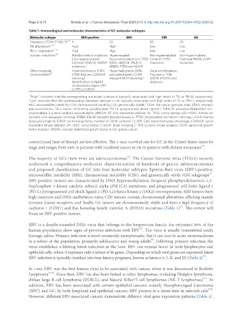
Table 1. Immunological and molecular characteristics of GC molecular subtypes
Molecular subtype EBV-positive MSI CIN GS
Frequency (TCGA STAD) % [4] 9 22 50 20
TIL abundance [5,6]* High High Low Low
PD-L1 expression [4-6]* High High Low Low
Somatic mutations [4] Standard rate of mutations Hypermutated Non-hypermutated Non-hypermutated
(non-hypermutated) Recurrent mutations in TP53, Common TP53 Common RHOA, CDH1
Common PI3KCA, ARID1A KRAS, ARID1A, PIK3CA, mutations mutations
mutations ERBB3, PTEN and HLA-B
Other molecular Hypermethylation (EBV- Hypermethylation (MSI- Gene amplifications,
characteristics [4] CIMP, frequent CDKN2A associated gastric CIMP, frequent in TKRs
silencing) frequent MLH1 silencing) (EGFR, VEGFA) and
Amplification in 9p24.1 deletions
chromosomal region (PD-
L1/PD-L2/JAK2)
*“High” indicates that the corresponding molecular subtype is typically associated with high levels of TIL or PD-L1, respectively.
“Low” indicates that the corresponding molecular subtype is not typically associated with high levels of TIL or PD-L1, respectively.
MSI: microsatellite instability; CIN: chromosomal instability; GS: genomically stable; TCGA: the cancer genome atlas; STAD: stomach
adenocarcinoma; TILs: tumor infiltrating lymphocytes; PD-L1: programmed death-ligand 1; PIK3CA: phosphatidylinositol-4,5-
bisphosphate 3-kinase catalytic subunit alpha; ARID1A: AT-rich interaction domain 1A; TP53: tumor protein p53; KRAS: Kirsten rat
sarcoma viral oncogene homolog; ERBB3: Erb-B2 receptor tyrosine kinase 3; PTEN: phosphatase and tensin homolog; HLA-B: human
leukocyte antigen B; RHOA: ras homolog family member A; CDH1: cadherin 1; CIMP: CpG island methylator phenotype; CDKN2A: cyclin
dependent kinase inhibitor 2A; JAK2: Janus kinase 2; MLH1: MutL homolog 1; TKR: tyrosine kinase receptor; EGFR: epidermal growth
factor receptor; VEGFA: vascular endothelial growth factor A; GC: gastric cancer
conventional lines of therapy are less effective. The 5-year survival rate for GC in the United States varies by
[2]
stage and ranges from 68% in patients with localized cancer to 5% in patients with distant metastases .
[3]
The majority of GCs (90%-95%) are adenocarcinomas . The Cancer Genome Atlas (TCGA) recently
undertook a comprehensive molecular characterization of hundreds of gastric adenocarcinomas
and proposed classification of GC into four molecular subtypes: Epstein-Barr virus (EBV)-positive,
[4]
microsatellite instability (MSI), chromosomal instability (CIN), and genomically stable (GS) subgroups .
EBV-positive tumors are characterized by DNA hypermethylation, frequent phosphatidylinositol-4,5-
bisphosphate 3-kinase catalytic subunit alpha (PIK3CA) mutations, and programmed cell death ligand 1
(PD-L1)/programmed cell death ligand 2 (PD-L2)/Janus kinase 2 (JAK2) overexpression; MSI tumors have
high mutation and DNA methylation rates; CIN tumors contain chromosomal alterations affecting mainly
tyrosine kinase receptors; and finally, GS tumors are chromosomally stable and have a high frequency of
[4]
cadherin 1 (CDH1) and Ras homolog family member A (RHOA) mutations [Table 1] . This review will
focus on EBV-positive tumors.
EBV is a double-stranded DNA virus that belongs to the herpesvirus family. An estimated 90% of the
[7]
human population show signs of previous infection with EBV . The virus is usually transmitted orally
through saliva. Primary infection is most commonly asymptomatic, but it can lead to acute mononucleosis
[8]
in a subset of the population, primarily adolescents and young adults . Following primary infection, the
virus establishes a lifelong latent infection in the host. EBV can remain latent in both lymphocytes and
epithelial cells, where it expresses only a subset of its genes. Depending on which viral genes are expressed, latent
EBV infection is typically classified into four latency programs, known as latency 0, I, II, and III [Table 2] .
[7]
In 1964, EBV was the first human virus to be associated with cancer, when it was discovered in Burkitt’s
Lymphoma [9,10] . Since then, EBV has also been linked to other lymphomas, including Hodgkin lymphoma,
[11]
diffuse large B-cell lymphoma (DLBCL), and Natural Killer/T-cell lymphomas (NK-T lymphomas) . In
addition, EBV has been associated with certain epithelial cancers, notably Nasopharyngeal Carcinoma
[11]
(NPC) and GC. In both lymphoid and epithelial cancers, EBV persists in a latent state in infected cells .
However, different EBV-associated cancers demonstrate different viral gene expression patterns [Table 2].