
Page 91 - Read Online
P. 91
Page 6 of 11 Kaufman et al. J Cancer Metastasis Treat 2019;5:73 I http://dx.doi.org/10.20517/2394-4722.2019.19
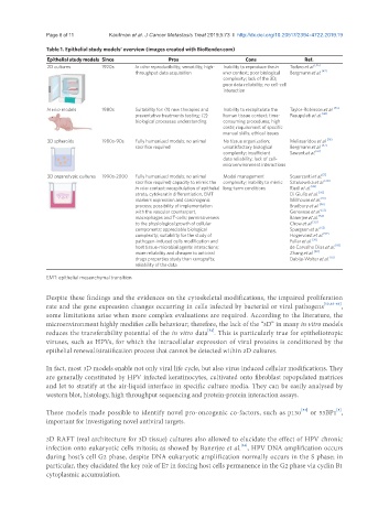
Table 1. Epithelial study models’ overview (images created with BioRender.com)
Epithelial study models Since Pros Cons Ref.
2D cultures 1920s In vitro reproducibility; versatility; high- Inability to reproduce the in Todaro et al. [45]
throughput data acquisition vivo context; poor biological Bergmann et al. [47]
complexity; lack of the 3D;
poor data reliability; no cell-cell
interaction
In vivo models 1980s Suitability for: (1) new therapies and Inability to recapitulate the Taylor-Robinson et al. [46]
preventative treatments testing; (2) human tissue context; time- Pasupuleti et al. [48]
biological processes understanding consuming procedures; high
costs; requirement of specific
manual skills; ethical issues
3D spheroids 1980s-90s Fully humanized models; no animal No tissue organization; Melissaridou et al. [16]
sacrifice required unsatisfactory biological Bergmann et al. [47]
complexity; insufficient Sawant et al. [61]
data reliability; lack of cell-
microenvironment interactions
3D organotypic cultures 1990s-2000 Fully humanized models; no animal Model management Squarzanti et al. [5]
sacrifice required; capacity to mimic the complexity; inability to mimic Sztukowska et al. [38]
in vivo context: recapitulation of epithelial long-term conditions Riedl et al. [49]
strata, cytokeratin differentiation, EMT Di Giulio et al. [50]
markers expression and carcinogenic Millhouse et al. [51]
process; possibility of implementation Bradbury et al. [56]
with the vascular counterpart, Genovese et al. [53]
macrophages and T-cells; permissiveness Banerjee et al. [54]
to the physiological growth of cellular Chow et al. [55]
components; appreciable biological Spurgeon et al. [52]
complexity; suitability for the study of Hogervorst et al. [57]
pathogen-induced cells modification and Fullar et al. [58]
host tissue-microbial agents interactions; de Carvalho Dias et al. [59]
more reliability and cheaper to antiviral Zhang et al. [60]
drugs properties study than xenografts; Dabija-Wolter et al. [62]
reliability of the data
EMT: epithelial mesenchymal transition
Despite these findings and the evidences on the cytoskeletal modifications, the impaired proliferation
rate and the gene expression changes occurring in cells infected by bacterial or viral pathogens [50,63-66] ,
some limitations arise when more complex evaluations are required. According to the literature, the
microenvironment highly modifies cells behaviour; therefore, the lack of the “3D” in many in vitro models
[52]
reduces the transferability potential of the in vitro data . This is particularly true for epitheliotropic
viruses, such as HPVs, for which the intracellular expression of viral proteins is conditioned by the
epithelial renewal/stratification process that cannot be detected within 2D cultures.
In fact, most 3D models enable not only viral life cycle, but also virus induced cellular modifications. They
are generally constituted by HPV infected keratinocytes, cultivated onto fibroblast repopulated matrices
and let to stratify at the air-liquid interface in specific culture media. They can be easily analysed by
western blot, histology, high throughput sequencing and protein-protein interaction assays.
[53]
These models made possible to identify novel pro-oncogenic co-factors, such as p130 or 53BP1 ,
[5]
important for investigating novel antiviral targets.
3D RAFT (real architecture for 3D tissue) cultures also allowed to elucidate the effect of HPV chronic
infection onto eukaryotic cells mitosis; as showed by Banerjee et al. , HPV DNA amplification occurs
[54]
during host’s cell G2 phase, despite DNA eukaryotic amplification normally occurs in the S phase; in
particular, they elucidated the key role of E7 in forcing host cells permanence in the G2 phase via cyclin B1
cytoplasmic accumulation.