
Page 94 - Read Online
P. 94
Vander Borght et al. J Cancer Metastasis Treat 2019;5:57 I http://dx.doi.org/10.20517/2394-4722.2019.0010 Page 3 of 6
Table 1. Neuroendocrine tumors of the lung
Name Smoking-associated % of all cases of lung cancer Overall Survival 5 y Mitotic index* Ki-67 index #
SCLC Yes 15-20 < 5% Median = 80 50%-100%
LCNEC Yes 3 15%-57% Median = 70 40%-80%
TC No 1-2 92%-100% < 2 < 5%
AC No 0.1-0.2 61%-68% 2-10 < 20%
#
Data from Ref.[6,8]. *Measure for rate of cell division; Ki-67 is a marker protein for cell division. SCLC: small cell lung cancer; LCNEC:
large cell neuroendocrine carcinoma; TC: typical carcinoid; AC: atypical carcinoid.
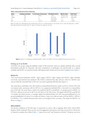
Figure 1. Reaction of monoclonal antibodies MUM-1, MUM-4 an MUM-6 with (truncated) E18 measured by an ELISA
Setting up of an ELISA
An ELISA was set up, using one antibody coated to the microtiter wells as a capture antibody and a second
biotinylated antibody for detection. The best combination of antibodies was MUMi-21B2 as a capture
antibody and MUM-6 as second antibody. Full experimental details can be found in Supplementary Figure 1.
RESULTS
Three monoclonal antibodies, MUM-1 (IgG1 kappa), MUM-4 (IgG1 kappa) and MUM-6 (IgG1 lambda),
were fully characterized and produced. The ELISA confirmed that they all react with E18. MUM-6 also
reacted with the truncated form of E18 that was used to generate MUMi-21B2 [Figure 1].
The monoclonal antibodies were also tested in immunohistochemistry assays on SCLC cell line NCI-H82
and control colon carcinoma cell line HCT116. In comparison antibody RNL-1, directed to an extracellular
part of NCAM, was used. Figure 2 shows the results for MUM-4 and MUM-6. A strong reaction of RNL-1 to
NCI-H82 cells an no reaction to HCT116 cells that lack NCAM is seen. The signal with MUM-4 and MUM-
6 is weaker but clearly present. A stronger signal was obtained when the cells were permeabilized with 4%
paraformaldehyde/0.5% triton X-100 (data not shown). Intact cells did not stain with the MUM-1, MUM-4
and MUM-6. This could be demonstrated most convincingly by flow cytometry. The results for MUM-1 are
shown in Figure 3.
DISCUSSION
The further validation of NCAM exon 18 expression in cancer cells is ongoing. More SCLC and LCNEC
biopsies will be analyzed. These are the two most aggressive neuroendocrine lung cancers, but it will be also
of interest to analyze the milder [Table 1] “typical carcinoid” and “atypical carcinoid”. However, our focus