
Page 231 - Read Online
P. 231
Giménez et al. J Cancer Metastasis Treat 2019;5:28 I http://dx.doi.org/10.20517/2394-4722.2018.75 Page 3 of 5
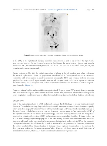
Figure 2. Positron emission tomography computer tomography showing multiple metastatic disease
in the UOQ of the right breast. Surgical treatment was determined and 43 out of 45 of the right ALND
were positive, seven of them with capsular rupture. In addition, the intramammary lymph node was also
positive for IDC, TN immunophenotype with a Ki67 of 63%. ASC and RT to the whole breast, axillary and
supraclavicular region was decided.
During controls, in May 2018 the patient complained of a lump in the left inguinal area, when performing
the physical exploration, a three cm lymph node was identified. A CNB reported metastatic carcinoma
compatible with breast cancer. A positron emission tomography (PET) scan revealed multiple abnormal
lymph nodes in the cervical, supraclavicular, mediastinal, retroperitoneal and inguinal regions. In addition,
two bone lesions, one in the pelvis and another in the humeral bone were described as well as unspecific
pleural nodules [Figure 2].
Treatment with carboplatin and gemcitabine was administered. However, a new PET revealed disease progression
with new muscular, hepatic, subcutaneous and bone lesions. The patient was admitted to the hospital for
severe respiratory insufficiency due to bilateral pleural effusion; finally, she died on October 10th of 2018.
DISCUSSION
One of the main explanations of CAM is aberrant drainage due to blockage of normal lymphatic routes.
[5]
Perre et al. described this theory; they studied 23 patients with breast cancer who underwent lymphoscintigraphy
before and after surgical treatment with or without radiotherapy. Only one patient presented drainage to
the contralateral axilla before treatment; however, this same event presented in four patients after ALND of
[6]
the opposite axilla and in one patient after radiotherapy. In a more recent publication, van der Ploeg et al.
observed, in patients with previous ALND for breast carcinoma, contralateral axillary drainage in four out
of 12 of them, during lymphoscintigraphy and SLNB. This finding becomes more relevant because two of the
four involved lymph nodes were positive for metastasis. Both patients underwent ALND on the same side
and reported alive and free of disease for 22 and 36 months after the procedure. In these two publications,
the authors concluded that this mechanism of drainage reflects regional dissemination to the node on the
direct pathway, making the “crossover metastasis” effect. However, a different outcome would be in cases of
untreated breast cancer, where CAM means dissemination beyond the regional nodes.