
Page 230 - Read Online
P. 230
Page 2 of 5 Giménez et al. J Cancer Metastasis Treat 2019;5:28 I http://dx.doi.org/10.20517/2394-4722.2018.75
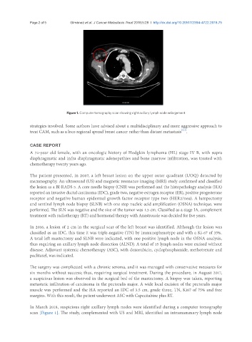
Figure 1. Computer tomography scan showing right axillary lymph node enlargement
strategies involved. Some authors have advised about a multidisciplinary and more aggressive approach to
[3,4]
treat CAM, such as a loco regional spread breast cancer rather than distant metastasis .
CASE REPORT
A 74-year old female, with an oncologic history of Hodgkin lymphoma (HL) stage IV B, with supra
diaphragmatic and infra diaphragmatic adenopathies and bone marrow infiltration, was treated with
chemotherapy twenty years ago.
The patient presented, in 2007, a left breast lesion on the upper outer quadrant (UOQ) detected by
mammography. An ultrasound (US) and magnetic resonance imaging (MRI) study confirmed and classified
the lesion as a BI-RADS 5. A core needle biopsy (CNB) was performed and the histopathology analysis (HA)
reported an invasive ductal carcinoma (IDC), grade two, negative estrogen receptor (ER), positive progesterone
receptor and negative human epidermal growth factor receptor type two (HER2/neu). A lumpectomy
and sentinel lymph node biopsy (SLNB) with one step nucleic acid amplification (OSNA) technique, were
performed. The SLN was negative and the size of the tumor was 1.5 cm. Classified as a stage IA, complement
treatment with radiotherapy (RT) and hormonal therapy with Anastrozole was decided for five years.
In 2016, a lesion of 2 cm in the surgical scar of the left breast was identified. Although the lesion was
classified as an IDC, this time it was triple-negative (TN) by immunophenotype and with a Ki-67 of 35%.
A total left mastectomy and SLNB were indicated, with one positive lymph node in the OSNA analysis,
thus requiring an axillary lymph node dissection (ALND). A total of 15 lymph nodes were excised without
disease. Adjuvant systemic chemotherapy (ASC), with doxorubicin, cyclophosphamide, methotrexate and
paclitaxel, was indicated.
The surgery was complicated with a chronic seroma, and it was managed with conservative measures for
six months without success; thus, requiring surgical treatment. During the procedure, in August 2017,
a suspicious lesion was observed in the surgical bed of the mastectomy. A biopsy was taken, reporting
metastatic infiltration of carcinoma in the pectoralis major. A wide local excision of the pectoralis major
muscle was performed and the HA reported an IDC of 3.5 cm, grade three, TN, Ki67 of 75% and free
margins. With this result, the patient underwent ASC with Capecitabine plus RT.
In March 2018, suspicious right axillary lymph nodes were identified during a computer tomography
scan [Figure 1]. The study, complemented with US and MRI, identified an intramammary lymph node