
Page 24 - Read Online
P. 24
Page 2 of 25 Kondapuram et al. J Cancer Metastasis Treat 2019;5:32 I http://dx.doi.org/10.20517/2394-4722.2018.105
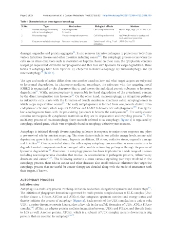
Table 1. Characteristics of three types of autophagy
Sl. No. Type Mechanism Biological effects Markers
1 Macroautophagy(commonly Autophagosome Cell killing and survival Beclin1, Atg5, Atg12, LC3-I and LC3-
referred as autophagy) formation II
2 Microautophagy Vacuole invagination process Cell killing and survival Hsc70 multi-vesicular bodies and
multi-vesicular lysosomes
3 Chaperon mediated autophagy Receptor mediated process Selective cell killing, T-cell LAMP-2A, Hsc70
activation
[1]
damaged organelles and protein aggregates . It also removes intrusive pathogens to protect our body from
[2,3]
various infectious diseases and other disorders including cancer . The autophagic process occurs when the
cells are in stress conditions such as starvation or hypoxia. Based on these cues, the cytoplasmic contents
(cargo) get sequestered within the autophagosome and then fuse with lysosome for cargo degradation. Three
forms of autophagy have been reported: (1) chaperon mediated autophagy; (2) microautophagy; and (3)
[4]
macroautophagy [Table 1].
The type and mode of action differs from one another based on how and what target cargoes are subjected
to lysosomal degradation. In chaperone mediated autophagy, the substrate with the targeting motif
KFERQ is recognized by the chaperone Hsc70, and moves the individual protein substrate to lysosome
[5]
degradation . While, microautophagy is responsible for basal degradation of the cytoplasmic content
[6]
by the direct invagination into lysosome . On the other hand, macroautophagy, an ubiquitous pathway
in eukaryotic cells, starts with the formation of double membrane structures called autophagosomes in
[3]
which cargo sequestration occurs . The early autophagosome is formed from components derived from
[7,8]
endoplasmic reticulum, which acquires V-ATPase and LAMP to become late autophagosome . Finally, the
[9]
late autophagosome fuses with the pre-existing lysosomes to become the autolysosome . The autolysosome
contains unrecognizable cytoplasmic materials as they are in degradation and recycling process . The
[10]
multi-step process of macroautophagy (here onwards referred to as autophagy; Figure 1) is regulated by
autophagy related genes, which were originally found in autophagy defective yeast mutants.
Autophagy is initiated through diverse signaling pathways in response to major stress response and plays
a pro-survival role by nutrient recycling. The stress factors include low cellular energy levels, amino acid
deprivation, growth factor withdrawal, hypoxia conditions, ER stress, oxidative stress, organelle damage
[11]
and infection . Over a period of stress, the cells employ autophagy process either to move contents or to
degrade harmful components such as damaged mitochondria or invading pathogens through the process of
[12]
lysosomal degradation . Aberration in autophagy process has been implicated in a wide range of diseases
including neurodegenerative disorders that involve the accumulation of pathogenic proteins, inflammatory
disorders and cancer [4,13] . The following sections discuss various signaling pathways involved in the
autophagy process, their role in cancer and other diseases; also small molecule inhibitors that target the
autophagy process that are useful for cancer therapy are detailed along with the mode of interaction with
their targets, if known.
AUTOPHAGY PROCESS
Initiation step
Autophagy is a multi-step process involving, initiation, nucleation, elongation/expansion and closure steps .
[14]
The initiation of phagophore formation is governed by multi-protein complex known as ULK complex (Unc-
51-like kinase 1, FIP200, ATG101 and ATG13), that integrates upstream nutrient and energy status and
thereby initiates the process of autophagy [Figure 2]. Each protein of the ULK complex has a unique role;
ULK1, a serine-threonine protein kinase, plays a key role in the scaffold formation of ULK1-ATG13-FIP200
[15]
complex ; ATG13, an adaptor protein mediates interaction between ULK1 and FIP200, and directly binds
to LC3 as well. Another protein, ATG101 which is a subunit of ULK complex recruits downstream Atg
proteins that are essential for autophagy [16,17] .