
Page 183 - Read Online
P. 183
Page 2 of 8 Hu et al. J Cancer Metastasis Treat 2018;4:39 I http://dx.doi.org/10.20517/2394-4722.2018.08
Gastric wall
Cancer cells
Peritoneum
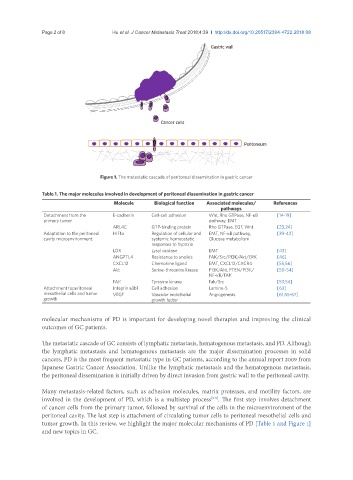
Figure 1. The metastatic cascade of peritoneal dissemination in gastric cancer
Table 1. The major molecules involved in development of peritoneal dissemination in gastric cancer
Molecule Biological function Associated molecules/ References
pathways
Detachment from the E-cadherin Cell-cell adhesion Wnt, Rho GTPase, NF-κB [14-19]
primary tumor pathway, EMT
ARL4C GTP-binding protein Rho GTPase, EGF, Wnt [23,24]
Adaptation to the peritoneal HIF1α Regulation of cellular and EMT, NF-κB pathway, [39-42]
cavity microenvironment systemic homeostatic Glucose metabolism
responses to hypoxia
LOX Lysyl oxidase EMT [43]
ANGPTL4 Resistance to anoikis FAK/Src/PI3K/Akt/ERK [46]
CXCL12 Chemokine ligand EMT, CXCL12/CXCR4 [55,56]
Akt Serine-threonine kinase PI3K/Akt, PTEN/PI3K/ [50-54]
NF-κB/FAK
FAK Tyrosine kinase Fak/Src [53,54]
Attachment toperitoneal Integrin a3b1 Cell adhesion Lamine-5 [63]
mesothelial cells and tumor VEGF Vascular endothelial Angiogenesis [61,65-67]
growth growth factor
molecular mechanisms of PD is important for developing novel therapies and improving the clinical
outcomes of GC patients.
The metastatic cascade of GC consists of lymphatic metastasis, hematogenous metastasis, and PD. Although
the lymphatic metastasis and hematogenous metastasis are the major dissemination processes in solid
cancers, PD is the most frequent metastatic type in GC patients, according to the annual report 2009 from
Japanese Gastric Cancer Association. Unlike the lymphatic metastasis and the hematogenous metastasis,
the peritoneal dissemination is initially driven by direct invasion from gastric wall to the peritoneal cavity.
Many metastasis-related factors, such as adhesion molecules, matrix proteases, and motility factors, are
involved in the development of PD, which is a multistep process . The first step involves detachment
[5-9]
of cancer cells from the primary tumor, followed by survival of the cells in the microenvironment of the
peritoneal cavity. The last step is attachment of circulating tumor cells to peritoneal mesothelial cells and
tumor growth. In this review, we highlight the major molecular mechanisms of PD [Table 1 and Figure 1]
and new topics in GC.