
Page 913 - Read Online
P. 913
Page 10 of 22 Mantovani et al. Hepatoma Res 2020;6:78 I http://dx.doi.org/10.20517/2394-5079.2020.75
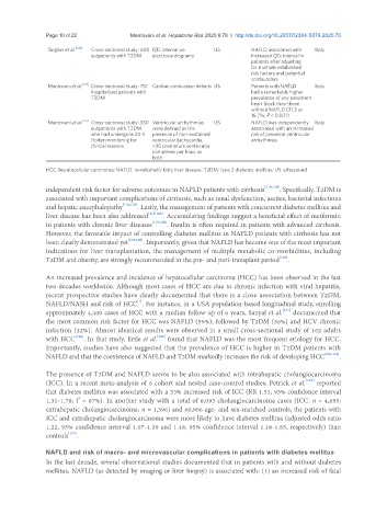
Targher et al. [126] Cross-sectional study: 400 QTc interval on US NAFLD associated with Italy
outpatients with T2DM electrocardiograms increased QTc interval in
patients after adjusting
for multiple established
risk factors and potential
confounders
Mantovani et al. [127] Cross-sectional study: 751 Cardiac conduction defects US Patients with NAFLD Italy
hospitalized patients with had a remarkably higher
T2DM prevalence of any persistent
heart block than those
without NAFLD (31.3 vs.
16.7%, P < 0.001)
Mantovani et al. [56] Cross-sectional study: 330 Ventricular arrhythmias US NAFLD was independently Italy
outpatients with T2DM were defined as the associated with an increased
who had undergone 24-h presence of non-sustained risk of prevalent ventricular
Holter monitoring for ventricular tachycardia, arrhythmias
clinical reasons >30 premature ventricular
complexes per hour, or
both
HCC: hepatocellular carcinoma; NAFLD: nonalcoholic fatty liver disease; T2DM: type 2 diabetes mellitus; US: ultrasound
independent risk factor for adverse outcomes in NAFLD patients with cirrhosis [119,120] . Specifically, T2DM is
associated with important complications of cirrhosis, such as renal dysfunction, ascites, bacterial infections
and hepatic encephalopathy [119,120] . Lastly, the management of patients with concurrent diabetes mellitus and
liver disease has been also addressed [119,120] . Accumulating findings suggest a beneficial effect of metformin
in patients with chronic liver diseases [119,120] . Insulin is often required in patients with advanced cirrhosis.
However, the favorable impact of controlling diabetes mellitus in NAFLD patients with cirrhosis has not
been clearly demonstrated yet [119,120] . Importantly, given that NAFLD has become one of the most important
indications for liver transplantation, the management of multiple metabolic co-morbidities, including
T2DM and obesity, are strongly recommended in the pre- and peri-transplant period [128] .
An increased prevalence and incidence of hepatocellular carcinoma (HCC) has been observed in the last
two decades worldwide. Although most cases of HCC are due to chronic infection with viral hepatitis,
recent prospective studies have clearly documented that there is a close association between T2DM,
[1]
NAFLD/NASH and risk of HCC . For instance, in a USA population-based longitudinal study, enrolling
approximately 4,400 cases of HCC with a median follow-up of 6 years, Sanyal et al. [121] documented that
the most common risk factor for HCC was NAFLD (59%), followed by T2DM (36%) and HCV chronic
infection (22%). Almost identical results were observed in a small cross-sectional study of 162 adults
with HCC [122] . In that study, Ertle et al. [122] found that NAFLD was the most frequent etiology for HCC.
Importantly, studies have also suggested that the prevalence of HCC is higher in T2DM patients with
NAFLD and that the coexistence of NAFLD and T2DM markedly increases the risk of developing HCC [129-131] .
The presence of T2DM and NAFLD seems to be also associated with intrahepatic cholangiocarcinoma
(ICC). In a recent meta-analysis of 6 cohort and nested case-control studies, Petrick et al. [132] reported
that diabetes mellitus was associated with a 53% increased risk of ICC (RR 1.53, 95% confidence interval
1.31-1.78; I = 67%). In another study with a total of 6,093 cholangiocarcinoma cases (ICC: n = 4,695;
2
extrahepatic cholangiocarcinoma: n = 1,396) and 60,906 age- and sex-matched controls, the patients with
ICC and extrahepatic cholangiocarcinoma were more likely to have diabetes mellitus (adjusted odds ratio
1.22, 95% confidence interval 1.07-1.39 and 1.48, 95% confidence interval 1.18-1.85, respectively) than
controls [133] .
NAFLD and risk of macro- and microvascular complications in patients with diabetes mellitus
In the last decade, several observational studies documented that in patients with and without diabetes
mellitus, NAFLD (as detected by imaging or liver biopsy) is associated with: (1) an increased risk of fatal