
Page 556 - Read Online
P. 556
Page 6 of 10 Bieu et al. Hepatoma Res 2020;6:49 I http://dx.doi.org/10.20517/2394-5079.2020.39
A
B
C
D
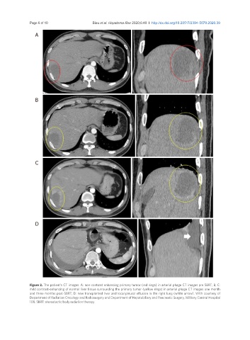
Figure 2. The patient’s CT images. A: non-contrast enhancing primary tumor (red rings) in arterial phage CT images pre SBRT; B, C:
mild contrast-enhancing of normal liver tissue surrounding the primary tumor (yellow rings) in arterial phage CT images one month
and three months post-SBRT; D: new transplanted liver and local pleural effusion in the right lung (white arrow). With courtesy of
Department of Radiation Oncology and Radiosurgery and Department of Hepatobiliary and Pancreatic Surgery, Military Central Hospital
108. SBRT: stereotactic body radiation therapy