
Page 553 - Read Online
P. 553
Bieu et al. Hepatoma Res 2020;6:49 I http://dx.doi.org/10.20517/2394-5079.2020.39 Page 3 of 10
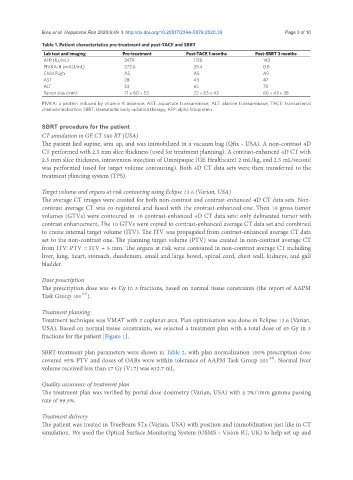
Table 1. Patient characteristics pre-treatment and post-TACE and SBRT
Lab test and imaging Pre-treatment Post-TACE 1 months Post-SBRT 3 months
AFP (IU/mL) 2479 1128 143
PIVKA-II (mAU/mL) 272.6 29.4 0.8
Child Pugh A5 A5 A5
AST 28 43 47
ALT 33 65 70
Tumor size (mm) 71 × 60 × 53 72 × 63 × 43 60 × 43 × 38
PIVKA: a protein induced by vitamin K absence; AST: aspartate transaminase; ALT: alanine transaminase; TACE: transarterial
chemoembolization; SBRT: stereotactic body radiation therapy; AFP: alpha fetoprotein
SBRT procedure for the patient
CT simulation in GE CT 580 RT (USA)
The patient lied supine, arm up, and was immobilized in a vacuum bag (Qfix - USA). A non-contrast 4D
CT performed with 2.5 mm slice thickness (used for treatment planning). A contrast-enhanced 4D CT with
2.5 mm slice thickness, intravenous injection of Omnipaque (GE Healthcare) 2 mL/kg, and 2.5 mL/second
was performed (used for target volume contouring). Both 4D CT data sets were then transferred to the
treatment planning system (TPS).
Target volume and organs at risk contouring using Eclipse 13.6 (Varian, USA)
The average CT images were created for both non-contrast and contrast-enhanced 4D CT data sets. Non-
contrast average CT was co-registered and fused with the contrast-enhanced one. Then 10 gross tumor
volumes (GTVs) were contoured in 10 contrast-enhanced 4D CT data sets: only delineated tumor with
contrast enhancement. The 10 GTVs were copied to contrast-enhanced average CT data set and combined
to create internal target volume (ITV). The ITV was propagated from contrast-enhanced average CT data
set to the non-contrast one. The planning target volume (PTV) was created in non-contrast average CT
from ITV: PTV = ITV + 5 mm. The organs at risk were contoured in non-contrast average CT including
liver, lung, heart, stomach, duodenum, small and large bowel, spinal cord, chest wall, kidneys, and gall
bladder.
Dose prescription
The prescription dose was 45 Gy in 3 fractions, based on normal tissue constraints (the report of AAPM
[17]
Task Group 101 ).
Treatment planning
Treatment technique was VMAT with 2 coplanar arcs. Plan optimization was done in Eclipse 13.6 (Varian,
USA). Based on normal tissue constraints, we selected a treatment plan with a total dose of 45 Gy in 3
fractions for the patient [Figure 1].
SBRT treatment plan parameters were shown in Table 2, with plan normalization 100% prescription dose
[16]
covered 95% PTV and doses of OARs were within tolerance of AAPM Task Group 101 . Normal liver
volume received less than 17 Gy (V17) was 812.7 mL.
Quality assurance of treatment plan
The treatment plan was verified by portal dose dosimetry (Varian, USA) with a 2%/1mm gamma passing
rate of 99.5%.
Treatment delivery
The patient was treated in TrueBeam STx (Varian, USA) with position and immobilization just like in CT
simulation. We used the Optical Surface Monitoring System (OSMS - Vision RT, UK) to help set up and