
Page 53 - Read Online
P. 53
Page 4 of 13 Méndez-Sánchez et al. Hepatoma Res 2020;6:5 I http://dx.doi.org/10.20517/2394-5079.2019.29
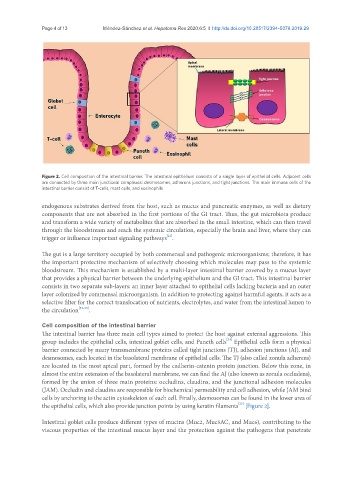
Figure 2. Cell composition of the intestinal barrier. The intestinal epithelium consists of a single layer of epithelial cells. Adjacent cells
are connected by three main junctional complexes: desmosomes, adherens junctions, and tight junctions. The main immune cells of the
intestinal barrier consist of T-cells, mast cells, and eosinophils
endogenous substrates derived from the host, such as mucus and pancreatic enzymes, as well as dietary
components that are not absorbed in the first portions of the GI tract. Thus, the gut microbiota produce
and transform a wide variety of metabolites that are absorbed in the small intestine, which can then travel
through the bloodstream and reach the systemic circulation, especially the brain and liver, where they can
[21]
trigger or influence important signaling pathways .
The gut is a large territory occupied by both commensal and pathogenic microorganisms; therefore, it has
the important protective mechanism of selectively choosing which molecules may pass to the systemic
bloodstream. This mechanism is established by a multi-layer intestinal barrier covered by a mucus layer
that provides a physical barrier between the underlying epithelium and the GI tract. This intestinal barrier
consists in two separate sub-layers: an inner layer attached to epithelial cells lacking bacteria and an outer
layer colonized by commensal microorganism. In addition to protecting against harmful agents, it acts as a
selective filter for the correct translocation of nutrients, electrolytes, and water from the intestinal lumen to
the circulation [22,23] .
Cell composition of the intestinal barrier
The intestinal barrier has three main cell types aimed to protect the host against external aggressions. This
[24]
group includes the epithelial cells, intestinal goblet cells, and Paneth cells Epithelial cells form a physical
barrier connected by many transmembrane proteins called tight junctions (TJ), adhesion junctions (AJ), and
desmosomes, each located in the basolateral membrane of epithelial cells. The TJ (also called zonula adherens)
are located in the most apical part, formed by the cadherin-catenin protein junction. Below this zone, in
almost the entire extension of the basolateral membrane, we can find the AJ (also known as zonula occludens),
formed by the union of three main proteins: occludins, claudins, and the junctional adhesion molecules
(JAM). Occludin and claudins are responsible for biochemical permeability and cell adhesion, while JAM bind
cells by anchoring to the actin cytoskeleton of each cell. Finally, desmosomes can be found in the lower area of
[23]
the epithelial cells, which also provide junction points by using keratin filaments [Figure 2].
Intestinal goblet cells produce different types of mucins (Muc2, Muc5AC, and Muc6), contributing to the
viscous properties of the intestinal mucus layer and the protection against the pathogens that penetrate