
Page 368 - Read Online
P. 368
Marasco et al. Hepatoma Res 2020;6:33 I http://dx.doi.org/10.20517/2394-5079.2020.01 Page 3 of 19
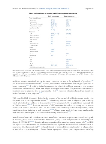
Table 1. Predictive factors for early and late HCC recurrence after liver resection
Early recurrence Late recurrence
Tumor related factors
Tumor size [27-29] [27-29]
Tumor grade [29-32] [29]
Macrovascular invasion [19,33-35] [19]
Microvascular invasion [19,36-39] [19,37]
Satellite nodules [40] -
Tumor-free margins [41-43] [41,43]
Biomarkers
AFP [32,35,44-46] [27,28]
Immunomarkers [47-54] -
ERASL-pre score [55] -
REACH score [56] -
SVR [57] [58-60]
HBV replication [61] [15,61]
Alcohol intake [62,63]
Others
MicroRNAs [64,65] -
Imaging factors [66-75]
IGC15 - [16]
Sarcopenia [76] -
NITs
LSM - [16,26,77]
SSM - [17]
FIB-4 - [78]
ALBI > 2 [55,79-82] [78,83,84]
Platelet/spleen length ratio - [17]
HCC: hepatocellular carcinoma; AFP: alpha-fetoprotein; ERASL: early recurrence after surgery for liver tumor; REACH-B: estimates risk of
HCC in patients with chronic hepatitis B; SVR: sustained virological response; HBV: hepatitis B virus; IGC15: indocyanine green retention
rate at 15 min; NITs: non-invasive tests; LSM: liver stiffness measurement; SSM: spleen stiffness measurement; FIB4: fibrosis-4 index;
ALBI: albumin-bilirubin grade
[28]
nodules ≥ 5 cm are associated with an increased recurrence rate due to the higher risk of portal vein
and micro-vascular invasion (MVI) . Besides, vascular invasion represents another good predictor of
[36]
tumor recurrence [19,27] . It could be defined as macroscopic, when it is visible on imaging or even on gross
examination, and microscopic, when seen only on histological examination. The presence of macrovascular
[19]
invasion is able to reduce the time to recurrence by 4-fold . Moreover, extension of portal vein thrombosis
is directly related to poor prognosis [33,34] .
With regard to MVI, it is usually defined as the presence of tumour emboli within the central hepatic vein,
[12]
the portal vein, or the large capsular vessels . Unfortunately, this evaluation is subject to great variability
[37]
which affects the true incidence of this condition . The presence of MVI is related to an increased risk
of HCC recurrence [19,38,39] . The main limitation of MVI assessment depends on its timing since it is often
obtained on resected specimens. MVI can be accurately assessed only on resected specimens, which
constitutes a strong limitation to such assessment [29,30,35] . Tumor grade (grade 3/3) and tumor size have also
been associated with early HCC recurrence and are strictly related to MVI [29-32] .
Several authors have tried to evaluate the usefulness of other pre-operative parameters beyond tumor grade
in predicting MVI, such as increased alpha-fetoprotein (AFP), L3-AFP and prothrombin induced by vit K
[85]
absence-II (PIVKA-II) [30,31,35] . Recently, a low concentration of the autophagy-related marker LC3 on HCC
and adjacent non-tumor tissues has also been found to be a significant predictor of both early and late HCC
recurrence. A further recent study evaluated the role of immunohistochemical markers in a large cohort
[47]
of resected HCC, concluding that 14 factors showed a prognostic role for predicting recurrence, including