
Page 257 - Read Online
P. 257
Bose et al. Hepatoma Res 2019;5:24 I http://dx.doi.org/10.20517/2394-5079.2019.10 Page 7 of 9
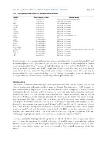
Table 3. Dysregulated lncRNAs observed in hepatocellular carcinoma
lncRNA Change in concentration Isolated sample
HULC Upregulation Hepatic colorectal metastasis samples
H19 Upregulation Malignant liver tissues
UCA1 Upregulation Liver tissues and serum
HOTAIR Upregulation Malignant liver tissues
MVIH Upregulation Malignant liver tissues
ATB Upregulation Malignant liver tissues
HOTTIP Upregulation Malignant liver tissues
MALAT-1 Upregulation Malignant liver tissues
VLDLR Upregulation Malignant liver tissues and EVs
TUC339 Upregulation EV
MEG3 Downregulation Malignant liver tissues
PTENP1 Downregulation Malignant liver tissues
DREH Downregulation Malignant liver tissues
WT1-AS Downregulation Malignant liver tissues
Uc002mbe.2 Downregulation Malignant liver tissues
XIST and FTX Downregulation Expressed more in liver malignant tissues of female than male
CPS1-IT1 Downregulation Malignant liver tissues
AOC-4P Downregulation Malignant liver tissues
HEIH Upregulation Malignant liver tissues
Recently, among various non-protein biomarkers, aberrant methylation, like hypomethylation in DNA and/
or hypermethylation in the CpG promoter gene, are found to be associated in the pathogenesis of different
tumors, including liver HCC [42,43] . A recent study identified a set of aberrantly methylated DNA markers
showing significant association with the HCC progression and interestingly, most of these hypermethylation
occur within the CpG domain . The identification of these circulating tumor DNAs carrying the
[44]
hypermethylated aberration within in the large cohort of HCC patients provides a promise of development
of a highly sensitive, noninvasive and accurate early detection platform for HCC.
CONCLUSION
HCC remains to be the most fatal malignant liver cancer worldwide even after the advances that has been
achieved in diagnostic and invasive medicine since last decades. The contemporary HCC treatment has
been intensive to early diagnosis and hepatic transplantation as medical management of this fatal disease.
Combination therapies performs well to downgrade the tumor and make it removable, that significantly
improve basic liver function and improve the timeline of survival, however, the early diagnosis has been the
most crucial deciding factor. In summary, informative feedback on the preclinical performance of image-
based techniques, AFP, AFP-L 3%, and DCP in the detection of HCC has created a vast and varied data set
since last few decades that serves as a fulcrum in the early diagnosis and medical management of HCC.
Recent observations demonstrate that many miRNAs and lncRNAs are differentially expressed in malignant
liver tissues, and their dysregulation as reflected in aberrant concentrations in various clinical samples
(tissues, serum, EVs etc.) were found to be correlated well with HCC progression, recurrence after liver
transplantation, chemoresistance etc. Thus, these miRNAs or lncRNAs may serve as potential biomarkers
for the diagnosis, prognosis, prediction of recurrence and therapeutic response of HCC .
[45]
However, a substantial heterogeneity among various cohorts of patients in terms of diagnostic criteria
leaves the space for evaluating the clinical performance of novel biomarkers, comprehensive studying
different variables associated with the malignant transformation in HCC and inclusion of some of the newer
biomarkers in surveillance strategy may prove to be crucial in future clinical management of this fatal
disease.