
Page 227 - Read Online
P. 227
Giorgio et al. Hepatoma Res 2019;5:20 I http://dx.doi.org/10.20517/2394-5079.2019.05 Page 3 of 9
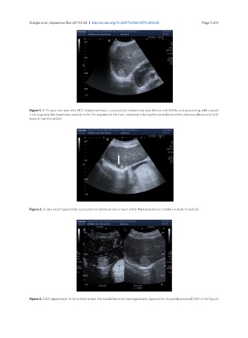
Figure 1. A 75-year old man with HCV related cirrhosis, successfully treated one year before with DAAs and presenting with a small
2 cm angioma-like hyperchoic nodule in the 7th segment of the liver, not present during the surveillance in the previous ultrasound (US)
exam 6 months before
Figure 2. A very small hypoechoic round portion (white arrow) is seen within the hyperechoic nodule ( nodule in nodule)
Figure 3. CEUS appearence: in the arterial phase, the nodule becomes homogeneously hyperechoic (hyperehancenced) (left of the figure)