
Page 329 - Read Online
P. 329
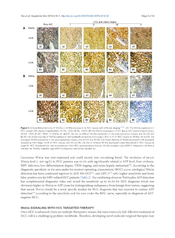
Yao et al. Hepatoma Res 2018;4:30  I  http://dx.doi.org/10.20517/2394-5079.2018.32                                                   Page 5 of 10

                                                                                      HCC and clinical stages
                               Para-HCC           I                                II                               III                                V
                 A  Wnt3a


                    ×200


                    ×400


                 B  Wnt5a


                    ×200


                    ×400


               Figure 1. Immunohistochemistry of Wnt3a or Wnt5a expression in HCC tissues with different staging [20,65] . (A) The Wnt3a expression in
               HCC tissues (SP, original magnification: A1-A5, ×200; B1-B5, ×400); (B) the Wnt5a expression in HCC tissue (SP, original magnification,
               A1-A5, ×200; B1-B5, ×400). In Wnt3a, A1 and B1, the low or without Wnt3a expression in the para-cancerous tissues, and A2-A5 and
               B2-B5, the brown staining of Wat3a expression with gradually increasing from stage I, II to III-IV of HCC tissues; In Wnt5a, A1 and B1, the
               strongest Wnt3a expression in the para-cancerous tissues, and A2-A4 and B2-B4, the brown staining of Wat3a expression with gradually
               decreasing from stage I to III of HCC tissues, and A5 and B5, the low or without Wnt5a expression were discovered in HCC tissues at
               stage IV. HCC: hepatocellular carcinoma tissues; Para-HCC: paracancerous tissues; Wnt3a: wingless-type MMTV integration site family
               member 3a; Wnt5a: wingless-type MMTV integration site family member 5a


               Cancerous Wnt3a was over-expressed and could secrete into circulating blood. The incidence of serum
               Wnt3a level (> 800 ng/L) in HCC patients was 92.5% with significantly related to AFP level, liver cirrhosis,
               HBV infection, low differentiation degree, TNM staging, and extra-hepatic metastasis . According to the
                                                                                         [19]
               diagnostic specificity or the area under the receiver operating characteristic (ROC) curve, serological Wnt3a
               detection has been confirmed superior to AFP, HS-GGT , and GPC-3  with higher sensitivity and lower
                                                               [72]
                                                                           [73]
               false-positive rate for HBV-related HCC patients [Table 2]. The combining of serum Wnt3a plus AFP detection
               has complemented diagnostic value and raised the sensitivity up to 96.3% for HCC diagnosis which was
               obviously higher in Wnt3a or AFP alone for distinguishing malignancy from benign liver lesions, suggesting
               that serum Wnt3a should be a novel specific marker for HCC diagnosis that was superior to routine AFP
               detection  according to the specificity and the area under the ROC curve, especially in diagnosis of AFP-
                       [74]
               negative HCC.


               Wnt3a SIGNALING WITH HCC TARGETED-THERAPY
               Once HCC is advanced, there are multiple therapeutic venues, but most eventually fail. Effective treatment of
               HCC still is a challenging problem worldwide. Therefore, developing novel molecule-targeted therapies may